Pectus Excavatum
What Is Pectus Excavatum (Funnel Chest)? Detailed Information on Symptoms, the Haller Index, the Vacuum Bell, and the Nuss and Ravitch Procedures.
Pectus Excavatum (Funnel Chest)
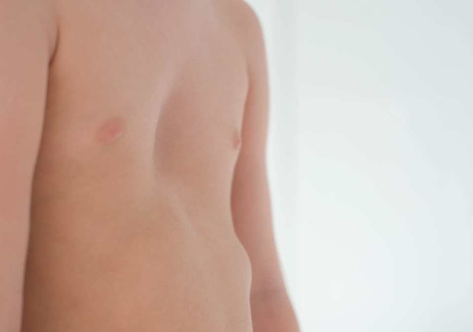
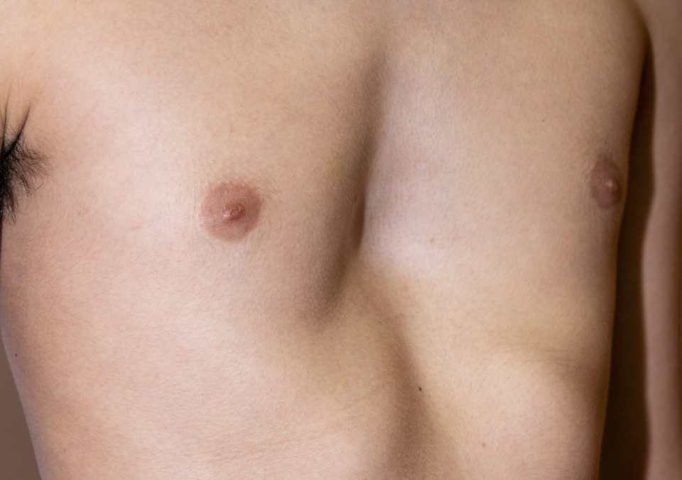
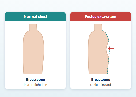
Pectus excavatum is a developmental chest wall deformity characterized by an inward, sunken appearance of the breastbone and the front of the chest wall. It is also known as "sunken chest" or "funnel chest."
The depression may be limited to the lower part of the breastbone, or it may involve a larger portion of the rib cage.
In some people the deformity is symmetric. In others, the breastbone (called the sternum) may be rotated, and the depression may be more pronounced on the right or left side.
Pectus excavatum may be present from birth. However, a deformity that is mild in childhood may become more noticeable, particularly during the rapid growth of adolescence.
Mild cases may cause only a difference in the appearance of the chest. In more pronounced deformities, the backward displacement of the sternum can reduce the anatomical space within the chest cavity and, in some people, may have functional effects on the heart and respiratory system, particularly during exercise.
Possible complaints include:
- Shortness of breath during exercise
- Tiring quickly
- Reduced exercise capacity
- Palpitations
- Chest pain or a feeling of pressure
- Postural changes
- Psychosocial effects related to body image
- Depth and width of the deformity
- Symmetry
- Sternal rotation
- Progression over time
- Anatomical effects on the heart and lungs
- Physical symptoms
- Exercise capacity
- Posture
- Age and growth status
- Body image and psychosocial effects
What Is Pectus Excavatum?
In a normal rib cage, the sternum sits at the front center of the chest. The ribs attach to the sternum through cartilage.
In pectus excavatum, the sternum and the adjacent costal cartilages appear displaced inward.
The deformity can take different anatomical forms:
- Limited to the lower part of the sternum
- Broad, involving the middle of the chest
- Extending to the lower rib cage
- Asymmetric, more pronounced on the right or left
- Accompanied by sternal rotation
- With outward flaring of the lower rib margins
What Causes Pectus Excavatum?
The exact mechanism by which pectus excavatum develops cannot be identified in every patient.
The deformity is thought to be related to the way the cartilage connecting the sternum and ribs grows and develops.
As the rib cage grows, structural differences in the development of the sternum, ribs, and costal cartilages may contribute to the inward shaping of the front chest wall.
Pectus excavatum is a developmental variation of the chest wall.
Sleeping position, carrying a heavy school bag, playing or not playing sports, poor nutrition, or anything the child or family did are not direct causes of pectus excavatum.
Poor posture does not cause the deformity either. However, rounded shoulders and a hunched back can make an existing chest depression look more pronounced.
Is Pectus Excavatum Hereditary?
Pectus excavatum may occur in more than one person in the same family.
Family members may have:
This suggests that genetic predisposition may play a role in some patients. However, the absence of a chest deformity in other family members does not mean that pectus excavatum will not develop. The type and severity of the deformity may also differ within the same family.
Family members may have:
- Pectus excavatum
- Pectus carinatum
- Rib cage asymmetries
- Scoliosis
- Joint hypermobility
- Certain connective tissue disorders
This suggests that genetic predisposition may play a role in some patients. However, the absence of a chest deformity in other family members does not mean that pectus excavatum will not develop. The type and severity of the deformity may also differ within the same family.
When Does Pectus Excavatum Become Noticeable?
Pectus excavatum may be present from birth.
In some children it is noticed at an early age, while in others it may not become apparent until the rib cage grows.
The deformity may become deeper or more noticeable, particularly during the rapid growth of adolescence.
During this period:
- The chest depression may deepen
- Sternal rotation may become more apparent
- Asymmetry between the right and left sides may increase
- The lower rib margins may flare outward
- The shoulders may round forward
- Postural changes may become more noticeable
- Exercise-related complaints may appear
- The impact on body image may increase
What Are the Symptoms of Pectus Excavatum?
In some people, pectus excavatum causes no physical complaints at all.
Symptoms may vary depending on the structure of the deformity, the position of the heart, the width of the rib cage, age, and level of physical activity.
Possible complaints include:
- Shortness of breath during exercise
- Tiring sooner than peers
- Reduced exercise capacity
- Difficulty running or climbing stairs
- Chest pain or a feeling of pressure
- Palpitations
- Being noticeably aware of one's heartbeat
- Dizziness
- A feeling of difficulty taking a deep breath
- Reduced performance in endurance activities
- Poor posture
- Back, neck, or shoulder pain
Can Pectus Excavatum Affect the Heart?
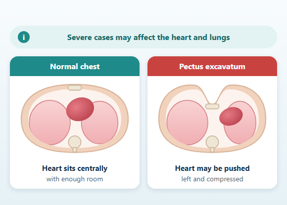
In severe pectus excavatum, the sternum may be displaced toward the space occupied by the heart.
The heart may appear pushed or rotated to the left. In some anatomical configurations, the right-sided chambers of the heart in particular may be compressed between the sternum and the spine.
In some patients, this anatomical change may affect the filling of the heart or its stroke volume, particularly during exercise.
As a result, symptoms such as the following may occur:
- Reduced exercise capacity
- Tiring quickly
- Palpitations
- Dizziness
- Chest discomfort
Does Pectus Excavatum Affect Exercise Capacity?
In some people, pectus excavatum may affect exercise capacity, particularly during strenuous physical activity.
While having no problems in daily life, a person may notice that they tire sooner than their peers during:
- Running
- Swimming
- Cycling
- Soccer
- Climbing stairs
- Endurance sports
Does Pectus Excavatum Affect Posture?
Some people with pectus excavatum may have:
- Rounded shoulders
- A rounded upper back
- A forward head posture
- Muscle imbalance around the shoulder blades
Can Pectus Excavatum Have Psychological Effects?
Pectus excavatum often becomes noticeable during childhood and adolescence, when body image is developing.
While some people are only minimally affected by the appearance of their chest, others may experience significant psychosocial difficulties.
A person may:
- Avoid changing clothes in front of others
- Not want to take part in swimming activities
- Stay away from beaches or pools
- Avoid tight-fitting clothing
- Not want to go to the gym
- Try to hide their chest in photographs
- Feel anxious in social or romantic relationships
- Withdraw from social settings
Conditions That May Accompany Pectus Excavatum
Pectus excavatum may occur on its own.
In some people, it may occur together with:
- Scoliosis
- Kyphosis
- Shoulder asymmetry
- Joint hypermobility
- Marfan syndrome
- Ehlers-Danlos syndrome
- Certain congenital heart conditions
- Certain changes in the heart valves or the structure of the aorta
Pectus Excavatum and Scoliosis
Pectus excavatum and scoliosis are distinct structural conditions, but they can occur together in the same person.
Shoulder and trunk asymmetry may be more noticeable, especially in asymmetric deformities with marked sternal rotation.
The examination may assess:
- Shoulder height
- Shoulder blades
- Spine
- Trunk rotation
- Right and left sides of the rib cage
How Is Pectus Excavatum Diagnosed?
The diagnosis is usually made through the medical history and physical examination.
The examination may assess:
- Location of the depression
- Deepest point
- Width of the deformity
- Symmetry
- Sternal rotation
- Flaring of the lower rib margins
- Flexibility of the chest wall
- Chest movement during breathing
- Shoulder and back posture
- Signs of scoliosis
- Scars from previous surgery or procedures
Which Tests May Be Used in Pectus Excavatum?
Tests are selected according to the patient's symptoms, the structure of the deformity, and the planned treatment.
When necessary, the following may be used:
The decision to operate is not based on the result of a single imaging study or test.
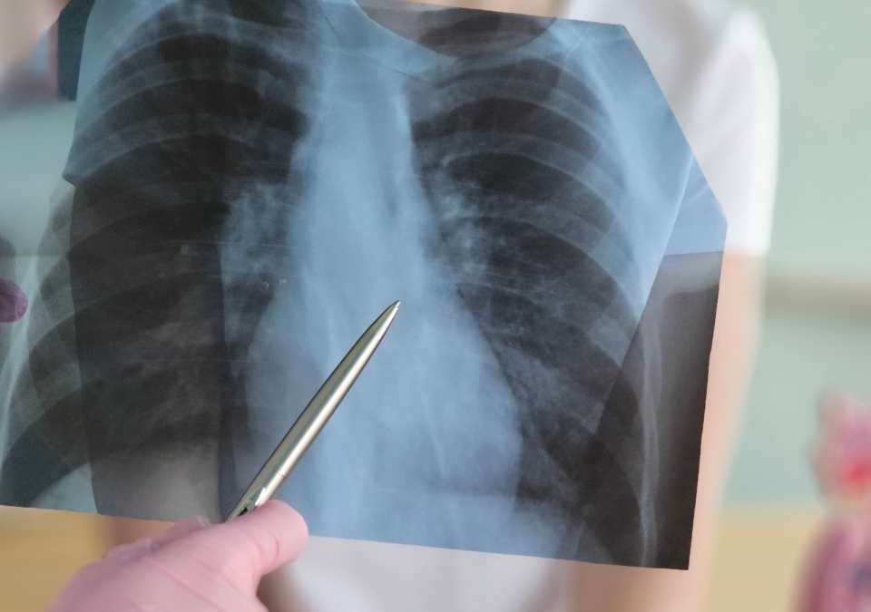
- Chest X-ray
- Computed tomography
- Magnetic resonance imaging
- Electrocardiography
- Echocardiography
- Pulmonary function tests
- Cardiopulmonary exercise testing
- Three-dimensional surface scanning
- Standardized photographic assessment
The decision to operate is not based on the result of a single imaging study or test.
What Does Computed Tomography Show?
Computed tomography can show the cross-sectional anatomy of the rib cage in detail.
When necessary, it can be used to assess:
Because computed tomography involves ionizing radiation, it should be used only when clinically necessary, especially in children and adolescents.
- Depth of the depression
- Narrowest point of the rib cage
- Sternal rotation
- Position of the heart
- Anatomical compression of the heart
- Rib cage asymmetry
- Haller index
- Correction index
- Anatomical features that affect surgical planning
Because computed tomography involves ionizing radiation, it should be used only when clinically necessary, especially in children and adolescents.
Can Magnetic Resonance Imaging Be Used?
Magnetic resonance imaging can help evaluate the chest and heart structures without using ionizing radiation.
MRI can be used to examine:
However, an MRI scan may take longer than a CT scan and may be more difficult to perform in some patients. The imaging method is chosen according to the patient's age, the clinical need, and the structure to be evaluated.
- Rib cage anatomy
- Position of the heart
- Compression of certain heart chambers
- Cardiac function
- Certain blood flow characteristics
However, an MRI scan may take longer than a CT scan and may be more difficult to perform in some patients. The imaging method is chosen according to the patient's age, the clinical need, and the structure to be evaluated.
What Is the Haller Index?
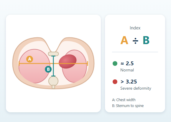
The Haller index is one of the measurements used to evaluate pectus excavatum on imaging.
It is calculated by dividing the widest horizontal diameter of the rib cage by the shortest front-to-back distance between the sternum and the spine.
The closer the sternum is to the spine, the higher the ratio.
Values of approximately 3.25 and above have historically been one of the commonly used thresholds for assessing a severe deformity.
However, the Haller index alone does not indicate the need for surgery.
Two people with the same Haller index may differ in:
For this reason, the Haller index is only one of many pieces of information in the clinical evaluation.
- Overall structure of the rib cage
- Asymmetry
- Position of the heart
- Cardiac compression
- Physical symptoms
- Exercise capacity
- Psychosocial effects
For this reason, the Haller index is only one of many pieces of information in the clinical evaluation.
What Is the Correction Index?
The correction index is one of the imaging measurements that expresses, as a percentage, the amount of correction needed for the sternum to reach a normal front-to-back position. In some rib cage configurations, it may provide information in addition to the Haller index. The correction index is not used on its own to make a surgical decision either. Imaging results are interpreted together with the physical examination, symptoms, cardiac evaluation, exercise capacity, and the course of the deformity over time.Why Is Echocardiography Performed?
Echocardiography uses sound waves to evaluate the structure and function of the heart. It may be used in the following situations:- Palpitations
- Chest pain during exercise
- Dizziness or fainting
- Heart murmur
- Suspected cardiac compression
- Suspected connective tissue disorder
- Preoperative evaluation
Pulmonary Function Tests
Pulmonary function tests provide information about airflow and breathing capacity of the lungs. They may be used especially in patients with shortness of breath or exercise-related complaints. In many people with pectus excavatum, pulmonary function tests performed at rest may be normal. The tests can also help distinguish asthma or another respiratory condition.What Is Cardiopulmonary Exercise Testing?
Cardiopulmonary exercise testing evaluates the combined response of the heart, lungs, and circulatory system during controlled physical activity. During the test, data such as the following may be examined:- Oxygen consumption
- Carbon dioxide production
- Heart rate
- Breathing response
- Workload
- Exercise duration
- Onset of symptoms
How Is the Severity of Pectus Excavatum Assessed?
Assessing severity is not just a matter of how deep the depression looks from the outside. The evaluation considers the following together:- Depth and width of the depression
- Symmetry
- Sternal rotation
- Haller and correction indices
- Position of the heart and possible compression
- Pulmonary function
- Exercise capacity
- Physical symptoms
- Progression of the deformity
- Posture
- Psychosocial effects
- Age and growth status
Does Pectus Excavatum Progress Over Time?
Pectus excavatum may become more noticeable, especially during periods of rapid growth. A depression that is mild in childhood may deepen or become more asymmetric during adolescence. The rate of progression varies from person to person. During follow-up, the following may be assessed:- Appearance of the chest
- Symmetry
- Growth
- Physical symptoms
- Exercise capacity
- Posture
- Scoliosis
- Psychosocial effects
Does Every Case of Pectus Excavatum Need Treatment?
No. Deformities that are mild, not progressing, and not significantly affecting the person's physical or psychosocial life can be monitored. Treatment may be considered in situations such as:- Progression of the deformity
- Reduced exercise capacity
- Significant effects on the heart or lungs
- Recurrent chest complaints
- Marked postural problems
- Impact on daily life or social activities
- Significant body image concerns
Can Exercise Correct Pectus Excavatum?
Exercise does not eliminate the structural cause of pectus excavatum. However, appropriate programs can:- Improve posture
- Bring the shoulders into a more balanced position
- Strengthen the back and shoulder muscles
- Increase the flexibility of the chest muscles
- Support breathing control
- Improve overall physical fitness
The Role of Physical Therapy in Pectus Excavatum
Physical therapy can provide support particularly for:- Rounded shoulders
- Hunched posture
- Weakness of the back muscles
- Reduced rib cage mobility
- Breathing control
- Physical preparation before surgery
- Regaining mobility after surgery
- Posture exercises
- Back and shoulder strengthening
- Stretching of the chest muscles
- Diaphragmatic breathing
- Rib cage mobility exercises
- General conditioning
What Is the Vacuum Bell?
The vacuum bell is a non-surgical device placed over the sunken area in pectus excavatum that creates negative pressure with the aim of lifting the sternum and the front chest wall forward. Once the device is placed on the chest, the air inside it is reduced in a controlled manner. The negative pressure created can cause the front chest wall to move forward. Vacuum bell treatment generally requires regular, long-term use. The response to treatment may be influenced by factors such as:- Age
- Chest wall flexibility
- Depth of the depression
- Symmetry of the deformity
- Consistency of use
- Total duration of treatment
Who May Be a Candidate for the Vacuum Bell?
The vacuum bell may be considered in selected patients, particularly those who:- Have a flexible chest wall
- Have a mild or moderate deformity
- Have a relatively symmetric depression
- Are able to commit to regular use of the device
How Is the Vacuum Bell Used?
There is no single universal schedule for vacuum bell use that applies to all patients. The duration of use and the negative pressure may be planned according to:- Age
- Chest wall flexibility
- Device type
- Structure of the deformity
- Skin tolerance
- Clinical response
What Are the Side Effects of the Vacuum Bell?
During or after vacuum bell use, the following may occur:- Skin redness
- Bruising
- Small visible blood vessels
- Tenderness
- Temporary swelling
- Chest discomfort
- Numbness
- Dizziness
Does the Vacuum Bell Provide Permanent Correction?
When the vacuum bell is applied, the sternum may temporarily move forward. This change, seen right after the device is removed, does not mean permanent remodeling. Regular, long-term use may be required for a permanent or lasting change. Significant improvement can be achieved in selected patients. However, the following cannot be guaranteed:- Complete correction
- Perfect symmetry
- A specific treatment duration
- No recurrence
- No need for surgery later
When Is Surgery Considered for Pectus Excavatum?
The decision to operate is not based solely on the appearance of the chest or the Haller index. Surgical treatment may be considered in selected patients in cases of:- Progressive deformity
- Significant physical complaints related to the deformity
- Significant cardiac compression on imaging
- Impaired cardiopulmonary function
- Markedly reduced exercise capacity
- Significant impact on daily life
- Serious and persistent psychosocial effects
- Non-surgical methods being unsuitable or not giving sufficient results
What Is the Right Age for Pectus Excavatum Surgery?
There is no single ideal age for surgery that applies to every patient. Timing is determined by taking into account:- Type and severity of the deformity
- Progression
- Chest wall flexibility
- Physical symptoms
- Cardiopulmonary effects
- Psychosocial status
- Whether growth is still ongoing
- Planned surgical technique
Can Pectus Excavatum Surgery Be Performed in Adults?
Yes. Pectus excavatum is not a condition treated only in children or adolescents. Surgical correction can also be performed in adults. However, because the chest wall is stiffer:- More force may be needed for correction
- More than one bar may be used
- Pain management may differ
- Recovery may take longer
- Surgical planning may be more detailed
- Age alone is not a barrier to surgery.
What Is the Nuss Procedure?
The Nuss procedure is a technique that aims to correct pectus excavatum using a minimally invasive surgical approach. One or more shaped metal bars are placed behind the sternum through small incisions on the sides of the chest. Once the bar is in the proper position, it supports the sternum forward and helps correct the sunken chest wall. The bars are left in the body for a certain period so that the rib cage can adapt to its new position, and are later removed in a separate surgical procedure. Although the Nuss procedure is performed through small skin incisions, it is a major chest wall correction procedure that is closely related to important anatomical structures, because a metal bar is passed behind the sternum.How Is the Nuss Procedure Performed?
The procedure is performed under general anesthesia. Although the surgical technique may vary according to the patient's anatomy, it may include the following steps:- Making incisions on the sides of the chest
- Viewing the chest cavity with a thoracoscopic camera
- Preparing a safe passage behind the sternum
- Shaping the metal bar to fit the chest
- Passing the bar behind the sternum
- Rotating the bar into position to lift the sternum
- Securing the bar to the chest wall
- Placing additional bars if needed
- Checking the chest cavity and the bar position
What Is a Nuss Bar?
A Nuss bar is a metal support shaped to fit the patient's rib cage. The bar:- Supports the sternum forward
- Helps maintain the corrected chest shape
- Provides mechanical support while the chest wall remodels
- More than one bar may be used depending on the width, stiffness, and asymmetry of the deformity.
How Long Does a Nuss Bar Stay In?
How long the bars remain in the body is not the same for all patients. Age, chest wall stiffness, the structure of the deformity, the correction achieved, and the surgical technique can affect the duration. The bars are generally left in place for several years to allow the rib cage to adapt to its new shape. The timing of bar removal is determined through regular clinical follow-up.How Is the Bar Removed?
Removal of the Nuss bar is a separate surgical procedure and is usually performed under general anesthesia. The previous side incisions may be used. After the tissues around the bar and fixation systems are freed, the metal bar is removed in a controlled manner. Although it may be a more limited procedure than the initial operation, it is not completely risk-free. Possible complications include:- Bleeding
- Pneumothorax
- Injury to the lung or pleura
- Infection
- Pain
- Technical difficulty due to bone or scar tissue around the bar
What Is the Ravitch Procedure?
The Ravitch method is based on correcting pectus excavatum with open surgery. During surgery, certain portions of the costal cartilages that contribute to the deformity may be reshaped or removed. When necessary, a corrective cut may be made in the sternum and the breastbone brought into a more forward position. The Ravitch method may be considered particularly for:- A very stiff chest wall
- Severe asymmetry
- A complex deformity
- Marked sternal rotation
- Some patients who have had previous pectus surgery
- Cases in which the Nuss method is not anatomically suitable
What Is the Difference Between the Nuss and Ravitch Procedures?
In the Nuss method, the breastbone is supported forward by one or more bars placed behind the sternum, without extensive removal of the costal cartilages. In the Ravitch method, the costal cartilages and, when necessary, the sternum are directly reshaped surgically. The Nuss method generally uses smaller side incisions, whereas the Ravitch method requires more direct surgical intervention on the front chest wall. Neither method can be said to be better than the other for all patients. The surgical approach is chosen by taking into account:- Anatomy of the deformity
- Age
- Chest wall flexibility
- Asymmetry
- Previous surgeries
- The patient's other medical characteristics
Evaluation Before Pectus Surgery
Before surgery, the following may be evaluated:- Onset and progression of the deformity
- Physical symptoms
- Exercise capacity
- Heart and lung status
- Scoliosis
- Signs of connective tissue disorder
- Previous surgeries
- Current medications
- Allergies
- Smoking
- Body image and expectations
- Support arrangements after surgery
Pain Control in Pectus Surgery
Because pectus excavatum surgery creates significant mechanical correction of the chest wall, postoperative pain management is an important part of treatment. Depending on the center and the patient, pain control may use methods such as:- Oral or intravenous pain medications
- Epidural analgesia
- Regional nerve blocks
- Paravertebral or erector spinae plane blocks
- Intercostal nerve cryoanalgesia
What Are the Risks of the Nuss Procedure?
The fact that the Nuss procedure is performed through small incisions does not mean it is risk-free. Possible complications include:- Bleeding
- Pneumothorax
- Hemothorax
- Pleural fluid accumulation
- Lung injury
- Rotation or displacement of the bar
- Problems related to fixation materials
- Infection
- Prolonged pain
- Nerve-related pain or sensory changes
- Metal reaction
- Atelectasis
- Lung infection
- Wound healing problems
- Insufficient correction
- Persistent asymmetry
- Recurrence of the deformity
- Need for repeat surgery
- Anesthesia-related complications
Bar Displacement
A Nuss bar exerts continuous mechanical force on the chest wall. Displacement or rotation of the bar may present with symptoms such as:- New or worsening chest pain
- A change in chest shape
- New asymmetry
- Shortness of breath
- Feeling the bar in a different position
Infection
Infection may develop at the incision site, around the implant, or in deeper tissues. Possible signs include:- Fever
- Redness
- Swelling
- Discharge
- Increasing pain
- Tenderness around the bar
- A change in general condition
Metal Allergy or Sensitivity
Reactions to metal implants are rare but may occur in some patients. People who have had significant reactions after contact with jewelry, watches, metal buttons, or previous implants should report this before surgery. In suspected cases, additional evaluation or different implant materials may be considered.Recovery After Pectus Surgery
In the early period, the following are important:- Pain control
- Breathing exercises
- Early mobilization
- Walking
- Assessment of the lungs
- Monitoring of the incision sites
- Checking the bar position
Activity Restrictions After Surgery
In the early period, the following may be temporarily restricted:- Heavy lifting
- Sudden twisting of the trunk
- Strenuous pushing or pulling
- Movements that put direct pressure on the chest
- Contact sports
- Activities with a high risk of falling
- Heavy weight training
Sports After Pectus Surgery
Return to sports is planned gradually. In the first stage, walking and light daily activities may be allowed. As recovery progresses, the following may be added to the program:- Light cardio
- Posture exercises
- Controlled stretching
- Light resistance strengthening
Posture and Physical Therapy After Surgery
Surgery can change the position of the sternum, but long-standing postural habits may not fully correct on their own. At the appropriate time after surgery, the following may be used:- Strengthening of the shoulder and back muscles
- Stretching of the chest muscles
- Posture work
- Diaphragmatic breathing
- Rib cage mobility exercises
- General conditioning
Returning to Work or School
The time it takes to return to school or work varies from person to person. The timing may be affected by:- Surgical method
- Pain level
- Number of bars used
- Physical demands of the job
- Commuting conditions
- Recovery
- Whether complications develop
Which Symptoms Should Be Evaluated After Surgery?
After surgery, medical evaluation may be needed in cases of:- Sudden or increasing chest pain
- Significant shortness of breath
- Fainting
- High fever
- Discharge from the incision site
- A sudden change in chest shape
- Suspicion that the bar has moved
- Newly developed noticeable asymmetry
- Uncontrollable pain
- A significant blow to the chest
Can Pectus Excavatum Recur After Treatment?
After vacuum bell or surgical treatment, there is a possibility that the deformity may become noticeable again. The risk of recurrence may be affected by factors such as:- Ongoing growth
- Structure of the chest wall
- Connective tissue disorders
- Severe asymmetry
- Insufficient correction
- Bar displacement
- Removal of the bar too early
- Adherence to treatment
Will the Chest Be Completely Symmetric After Surgery?
The goal of surgery is to reduce the deformity and bring the chest wall into a more balanced shape. However, perfect symmetry cannot be guaranteed. Some asymmetry may persist, particularly in people with:- Sternal rotation
- Rib asymmetry
- Scoliosis
- Differences in muscle development
- Previous surgeries
- Complex chest wall anatomy
Is Pectus Excavatum Surgery a Definitive Solution?
In suitable patients, surgical treatment can provide significant and long-lasting improvement in the shape of the chest wall. However, the following cannot be guaranteed:- Complete symmetry
- Resolution of all physical symptoms
- A definite increase in exercise capacity
- Complete resolution of pain
- Resolution of all body image concerns
- The deformity never recurring
- No complications
Follow-Up After Treatment
The follow-up process varies according to the treatment received. At follow-up visits, the following may be assessed:- Appearance of the chest wall
- Symmetry
- Persistence or recurrence of the deformity
- Bar position
- Incision sites
- Pain
- Respiratory complaints
- Exercise capacity
- Posture
- Scoliosis
- Return to daily life and sports
- Psychosocial status
- Timing of bar removal
When Is Medical Evaluation Needed?
An evaluation for pectus excavatum may be carried out in the following situations:- Rapid deepening of the chest depression
- Noticeable or increasing asymmetry
- Shortness of breath during exercise
- Recurrent chest pain
- Palpitations
- Dizziness or fainting
- A marked drop in performance compared with peers
- Scoliosis
- Body image concerns that affect daily life
- Considering vacuum bell or surgical treatment options
Frequently Asked Questions About Pectus Excavatum
Does pectus excavatum go away on its own?
The appearance of mild deformities may change during growth, but it should not be assumed that structural pectus excavatum will completely disappear on its own. In some children, the deformity may become more noticeable, particularly during the rapid growth of adolescence.
Is pectus excavatum dangerous?
In many people, pectus excavatum does not cause serious heart or lung problems. However, in severe deformities, cardiopulmonary effects may occur, particularly during exercise. The evaluation should go beyond the appearance of the deformity.
What Haller index value requires surgery?
A single Haller index value does not automatically mean that surgery is needed. Although values of approximately 3.25 and above are one of the commonly used criteria for assessing a severe deformity, symptoms, compression of the heart, exercise capacity, the structure of the deformity, and psychosocial effects are evaluated together.
Does pectus excavatum prevent playing sports?
Many people with pectus excavatum can be physically active without any sports restrictions. If there is significant shortness of breath, chest pain, palpitations, dizziness, or reduced performance during exercise, the cause should be evaluated separately.
Can exercise completely correct pectus excavatum?
Exercise can improve posture, muscle strength, and overall fitness, but it is not expected to completely correct the structural depression of the sternum and costal cartilages on its own.
In whom may the vacuum bell be more effective?
More favorable results may be achieved in younger patients with a more flexible chest wall and a mild or moderate, relatively symmetric deformity. Nevertheless, patient selection requires individual evaluation.
Can the vacuum bell replace surgery?
Not for every patient. The vacuum bell may be a non-surgical option for selected patients with pectus excavatum. Different treatments may be needed for deformities that are stiff, severe, markedly asymmetric, or associated with significant cardiopulmonary effects.
Why is a bar used in the Nuss procedure?
The metal bar supports the sternum forward and helps the chest wall stay in its corrected position. Once the rib cage has adapted to its new shape, the bar is removed in a separate surgical procedure.
Does the bar stay in for life after the Nuss procedure?
No. The bar is generally left in the body for a certain period and then removed. The exact duration varies according to age, chest wall characteristics, and the surgical plan.
Does pectus excavatum recur after surgery?
Recurrence is possible but does not occur in every patient. Ongoing growth, the structure of the deformity, connective tissue characteristics, the condition of the bar, and the stability of the correction achieved can affect the risk.
Conclusion
Pectus excavatum is a developmental chest wall deformity characterized by an inward, sunken appearance of the sternum and the front chest wall. Mild cases may cause only a difference in the appearance of the chest. In more severe deformities, displacement of the sternum toward the chest cavity may affect the position of the heart and cardiopulmonary function in some people. These effects may be more pronounced, particularly during strenuous physical activity. The evaluation of pectus excavatum is not based solely on outward appearance or the Haller index.The physical examination, the anatomy and progression of the deformity, cardiac evaluation, pulmonary function, exercise capacity, posture, and psychosocial effects are considered together. Not every patient with pectus excavatum needs surgery. For mild or stable deformities, observation, exercise, and physical therapy may be used. In selected patients with a flexible chest wall, the vacuum bell may be considered. For patients who need surgical treatment, the Nuss and Ravitch methods may be used.
In the Nuss method, one or more metal bars placed behind the sternum support the breastbone forward. In the Ravitch method, the costal cartilages and, when necessary, the sternum are directly reshaped.
Which method is appropriate is determined by the anatomy of the deformity, chest wall flexibility, age, previous treatments, and the patient's other clinical characteristics. The goal of treatment is not only to change the appearance of the chest. Physical complaints, heart and lung function, exercise capacity, body image, and overall quality of life should all be evaluated together.
Disclaimer: The information on this page is provided for general informational purposes. It does not replace a medical examination, diagnosis, or an individualized treatment plan. The evaluation and treatment options for pectus excavatum may vary depending on the person's age, chest wall anatomy, physical symptoms, cardiopulmonary findings, and other health characteristics.