Emphysema
What is emphysema? Symptoms, CT and DLCO Evaluation, Bullae, Bullectomy, Lung Volume Reduction, and Endobronchial Valve Treatments.
What Is Emphysema? Symptoms, Diagnosis, and Treatment
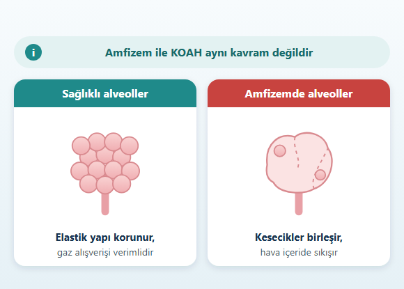
Emphysema is a chronic lung disease characterized by damage to the walls of the alveoli, the smallest air sacs of the lungs, and loss of the normal elastic structure of lung tissue.
As the alveolar walls are damaged, small air sacs can merge into larger spaces that are less efficient for gas exchange.
The elastic recoil of the lung tissue also decreases.
As a result:
- Breathing out may become harder
- Air may become trapped in the lungs
- The lungs may become overinflated
- The diaphragm may flatten
- The breathing muscles may work less efficiently
- The gas exchange surface may decrease
- Exercise capacity may decline
What Is Emphysema?
Healthy lungs contain millions of alveoli.
The thin walls of the alveoli are an important part of the structure that allows:
As a result of these changes:
The thin walls of the alveoli are an important part of the structure that allows:
- Oxygen to pass into the blood
- Carbon dioxide to be removed from the blood
- The lung to expand when breathing in
- The lung to recoil elastically when breathing out
As a result of these changes:
- Air spaces may enlarge
- The elastic recoil of lung tissue may decrease
- Small airways may close earlier during exhalation
- Air may become trapped in the lung
- Lung volume may increase
- The mechanical efficiency of the breathing muscles may decline
Are Emphysema and COPD the Same Disease?
No.
COPD is a broader clinical disease referring to persistent airflow limitation.
Emphysema describes structural damage to lung tissue.
In a COPD patient, the following may be present together in varying proportions:
Some people have emphysema-predominant COPD, while in others airway narrowing or sputum problems may be more prominent. For this reason, seeing emphysema on a CT scan does not mean the same thing as a diagnosis of COPD. Spirometry is required to confirm a diagnosis of COPD.
In a COPD patient, the following may be present together in varying proportions:
- Emphysema
- Small airway disease
- Chronic bronchitis
Some people have emphysema-predominant COPD, while in others airway narrowing or sputum problems may be more prominent. For this reason, seeing emphysema on a CT scan does not mean the same thing as a diagnosis of COPD. Spirometry is required to confirm a diagnosis of COPD.
What Causes Emphysema?
Emphysema can develop as a result of the interaction between damage to lung tissue caused by long-term harmful exposures and a person's genetic and biological susceptibility.
The main risk factors may include:
Smoking is one of the most important risk factors for emphysema, but the disease does not occur only in people who smoke.
The main risk factors may include:
- Smoking
- Other tobacco products
- Secondhand smoke exposure
- Occupational dusts
- Chemical fumes
- Biomass smoke
- Air pollution
- Certain long-term respiratory exposures
- Alpha-1 antitrypsin deficiency
- Inadequate lung development in some people
Smoking is one of the most important risk factors for emphysema, but the disease does not occur only in people who smoke.
Can Nonsmokers Develop Emphysema?
Yes.
Emphysema can also develop in people who have never smoked.
Possible causes may include:
In emphysema that develops at a young age, or that is more severe than expected based on smoking history, the underlying causes may need to be evaluated separately.
Possible causes may include:
- Alpha-1 antitrypsin deficiency
- Biomass smoke
- Occupational exposure
- Air pollution
- Previous lung damage
- Differences in lung development
- Other, less common causes
In emphysema that develops at a young age, or that is more severe than expected based on smoking history, the underlying causes may need to be evaluated separately.
What Is Alpha-1 Antitrypsin Deficiency?
Alpha-1 antitrypsin is a protein that helps protect lung tissue against the destructive effects of certain enzymes released during inflammation.
Inherited alpha-1 antitrypsin deficiency can make the lungs more prone to emphysema.
This deficiency is more likely to be considered in the following situations:
- Emphysema at a relatively young age
- Lung damage too advanced to be explained by smoking history
- Early-onset COPD or emphysema in the family
- Emphysema that is more pronounced in the lower parts of the lungs
- Unexplained liver disease
What Are the Types of Emphysema?
Emphysema can occur in different types depending on which part of the lung lobule is affected and how the damage is distributed.
Centrilobular Emphysema
Centrilobular, or centriacinar, emphysema is particularly associated with smoking. It begins in areas near the center of the lung lobule. It is often more pronounced in the upper parts of the lungs. The distribution of damage may be irregular.Panlobular Emphysema
In panlobular, or panacinar, emphysema, a larger part of the lung lobule is affected. It may be associated particularly with alpha-1 antitrypsin deficiency and may be more pronounced in the lower regions of the lungs.Paraseptal Emphysema
Paraseptal emphysema develops at the outer edges of the lungs and in areas close to the pleura. It may be associated with bulla formation and, in some people, with spontaneous pneumothorax.Irregular Emphysema
Irregular emphysema may be seen around areas of previous lung damage or fibrosis. More than one type of emphysema can be present in the same person.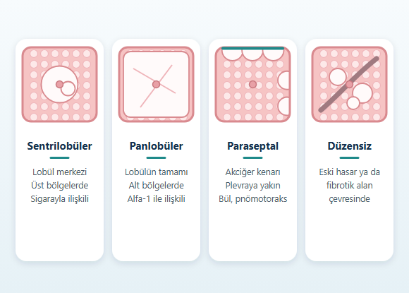
What Are the Symptoms of Emphysema?
One of the most common complaints in emphysema is progressive shortness of breath.
Possible symptoms may include:
If chronic bronchitis or other airway diseases are also present, cough and sputum may occur as well. In some patients in whom emphysema predominates, sputum is less prominent.
Possible symptoms may include:
- Shortness of breath on exertion
- Reduced walking capacity
- Difficulty climbing stairs
- Walking more slowly than peers
- Early fatigue during exercise
- A feeling of being unable to breathe out fully
- Prolonged exhalation
- A feeling of excessive fullness in the chest
- Reduced exercise capacity
- Weight loss in advanced disease
- More noticeable use of the accessory breathing muscles
If chronic bronchitis or other airway diseases are also present, cough and sputum may occur as well. In some patients in whom emphysema predominates, sputum is less prominent.
Why Does Emphysema Cause Shortness of Breath?
After a breath is taken, healthy lung tissue tends to recoil thanks to its elastic structure.
This recoil force helps push the air out.
In emphysema, this elastic support is reduced.
During exhalation, the small airways may narrow or close earlier.
As a result, air becomes trapped inside the lungs.
As air trapping increases:
As air trapping increases:
- Lung volume increases
- The diaphragm flattens
- The breathing muscles work at a mechanical disadvantage
- More energy is needed for each breath
- Exercise may become increasingly difficult
What Is Hyperinflation?
Hyperinflation is a condition in which more air than normal remains in the lungs and the lungs are overinflated.
It can develop in emphysema because air cannot be fully expelled during exhalation.
Static Hyperinflation
This is when the lungs remain at a higher-than-normal volume even at rest. It can be assessed with lung volume measurements such as residual volume and total lung capacity.Dynamic Hyperinflation
When breathing speeds up during exercise, a new breath may begin before the previous breath has been fully exhaled. With each breath, a little more air may build up in the lungs. This can cause marked shortness of breath, especially during physical activity.How Does Hyperinflation Affect the Diaphragm?
The diaphragm is normally a dome-shaped, curved muscle. Overinflation of the lungs can push the diaphragm downward and make it flatter. A flattened diaphragm may work with less mechanical advantage when breathing in. In this case, use of the accessory breathing muscles around the chest and neck may increase. One of the main goals of volume reduction methods in the treatment of advanced emphysema is to reduce this mechanical disadvantage.Does Emphysema Affect Oxygen Levels?
It can.
Loss of alveolar walls can reduce the surface area available for gas exchange.
In some patients, low oxygen may become more noticeable particularly:
Not every patient with emphysema needs to use oxygen. Oxygen therapy should be planned based on objective oxygen measurements, not on shortness of breath alone.
In some patients, low oxygen may become more noticeable particularly:
- During exercise
- During sleep
- During respiratory tract infections
- In the advanced stages of the disease
Not every patient with emphysema needs to use oxygen. Oxygen therapy should be planned based on objective oxygen measurements, not on shortness of breath alone.
Can Emphysema Cause Weight and Muscle Loss?
Weight and muscle loss can occur in advanced emphysema.
Reasons for this may include:
Muscle loss can further reduce exercise capacity and may affect the ability to tolerate advanced surgical or bronchoscopic procedures.
Reasons for this may include:
- Increased energy spent on breathing
- Reduced appetite
- Shortness of breath while eating
- Reduced physical activity
- Recurrent exacerbations
- Systemic effects of advanced chronic lung disease
Muscle loss can further reduce exercise capacity and may affect the ability to tolerate advanced surgical or bronchoscopic procedures.
How Is Emphysema Diagnosed?
A single test may not be sufficient to evaluate emphysema.
The evaluation may include tests such as:
Computed tomography is especially important for showing emphysema anatomically and evaluating its distribution.
The evaluation may include tests such as:
- Medical history
- Smoking and other exposures
- Physical examination
- Spirometry
- Lung volumes
- DLCO
- Oxygen measurement
- Arterial blood gas
- Computed tomography
- Exercise tests
Computed tomography is especially important for showing emphysema anatomically and evaluating its distribution.
Does Spirometry Show Emphysema?
Spirometry measures airflow limitation.
It does not directly show the amount of emphysematous tissue.
The key measurements are:
Even when emphysema is present on CT, spirometry may not yet show significant airflow limitation. Conversely, in a person with prominent small airway disease, spirometry may be severely impaired even though emphysema is limited. For this reason, spirometry and CT are not substitutes for each other but complementary tests.
The key measurements are:
- FEV1
- FVC
- FEV1/FVC
Even when emphysema is present on CT, spirometry may not yet show significant airflow limitation. Conversely, in a person with prominent small airway disease, spirometry may be severely impaired even though emphysema is limited. For this reason, spirometry and CT are not substitutes for each other but complementary tests.
What Does FEV1 Show in Emphysema?
FEV1 is the amount of air a person can exhale in the first second of a forced breath out.
It provides information about the degree of airflow limitation.
However, FEV1 alone does not show:
A more comprehensive physiological and anatomical evaluation is needed when advanced emphysema treatments are being considered.
However, FEV1 alone does not show:
- The amount of emphysema
- The distribution of emphysema
- The degree of hyperinflation
- Exercise capacity
- Suitability for lung volume reduction treatment
A more comprehensive physiological and anatomical evaluation is needed when advanced emphysema treatments are being considered.
What Is DLCO?
DLCO is a pulmonary function test that evaluates the diffusing capacity of the lungs.
It provides information about how effectively gases can pass from the alveoli into the blood.
Because the alveolar surface is reduced in emphysema, DLCO may decrease.
This test can help evaluate:
A low DLCO is not caused only by emphysema. Anemia, pulmonary fibrosis, and pulmonary vascular diseases can also reduce DLCO.
This test can help evaluate:
- Impairment of gas exchange
- The tendency for oxygen levels to drop during exercise
- Overall physiological reserve
- The risk of certain surgical procedures
A low DLCO is not caused only by emphysema. Anemia, pulmonary fibrosis, and pulmonary vascular diseases can also reduce DLCO.
What Is Body Plethysmography?
Body plethysmography can measure the air volumes in the lungs in detail.
In particular, the following can be evaluated:
In advanced emphysema, not only FEV1 but also how much air is trapped in the lungs is important for treatment planning.
In particular, the following can be evaluated:
- Residual volume
- Total lung capacity
- Functional residual capacity
- Air trapping
- Hyperinflation
In advanced emphysema, not only FEV1 but also how much air is trapped in the lungs is important for treatment planning.
Why Is Computed Tomography Important in Emphysema?
Computed tomography is one of the most important imaging methods for evaluating emphysema.
CT can be used to evaluate:
The importance of CT increases further when bronchoscopic or surgical lung volume reduction is being considered.
CT can be used to evaluate:
- The presence of emphysema
- The extent of damage
- Which lobes are most affected
- Upper- or lower-lobe predominance
- Homogeneous or heterogeneous distribution
- Bullae
- Bronchiectasis
- Lung nodules
- Other lung diseases
- Possible target areas for advanced treatment
The importance of CT increases further when bronchoscopic or surgical lung volume reduction is being considered.
What Is Quantitative CT Analysis?
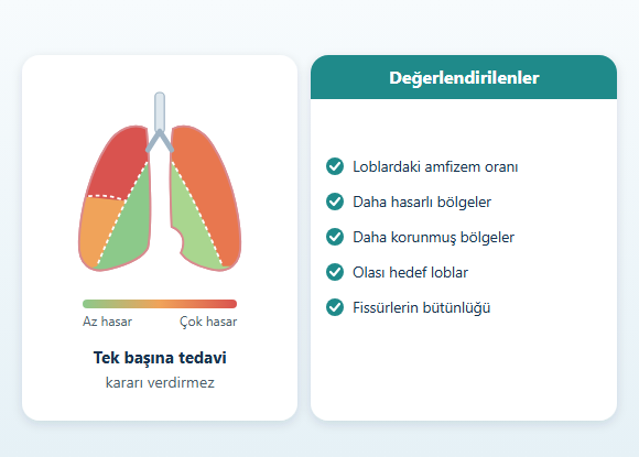
Computer-assisted CT analysis may be used in selected patients with advanced emphysema.
These systems can help evaluate:
- The proportion of emphysema in different lobes
- More severely damaged areas
- Better-preserved areas
- Potential target lobes
- The integrity of the fissures between the lobes
What Do Homogeneous and Heterogeneous Emphysema Mean?
In heterogeneous emphysema, some areas of the lung are markedly more damaged than others. In homogeneous emphysema, the damage is distributed more evenly throughout the lungs. This distinction is important for lung volume reduction methods. The basic principle of volume reduction is to target the areas that are most damaged and that contribute most to hyperinflation. Marked heterogeneous emphysema may be an advantage for certain treatments. However, selected patients with homogeneous emphysema may also be evaluated for certain advanced treatments.What Is a Bulla?
A bulla is the name given to a large air-filled space that develops as a result of emphysema. Small bullae may not cause any additional symptoms. Larger bullae, however, may:- Occupy a significant part of the chest cavity
- Compress better-functioning lung tissue
- Impair diaphragm movement
- Contribute to shortness of breath
- Become infected
- Be associated with pneumothorax
- Although a bulla and a pneumothorax are different conditions, distinguishing between them on imaging may be necessary.
What Is a Giant Bulla?
A giant bulla is a large emphysematous air space that occupies a significant part of the chest cavity. Its clinical significance is determined by:- The size of the bulla
- Its location
- The pressure it puts on surrounding lung tissue
- The quality of the remaining lung tissue
- The patient's symptoms
- A history of pneumothorax or infection
What Is Bullectomy?
Bullectomy is the surgical removal of large, nonfunctioning bullae. Surgical evaluation may be considered particularly when a large bulla:- Compresses better-functioning lung
- Occupies a significant part of the chest cavity
- Causes marked shortness of breath
- Is associated with recurrent pneumothorax
- Causes recurrent infections
What Are the Risks of Bullectomy?
Possible complications may include:- Prolonged air leak
- Pneumothorax
- Bleeding
- Infection
- Pneumonia
- Respiratory failure
- Heart rhythm problems
- Need for intensive care
- Reoperation
- Anesthesia complications
How Is Emphysema Treated?
Treatment is planned according to the severity of emphysema, symptoms, airflow limitation, hyperinflation, oxygen levels, and other health problems. Standard treatment steps may include:- Quitting smoking
- Appropriate inhaled bronchodilator treatments
- Other inhaled treatments in patients selected according to COPD characteristics
- Review of vaccinations
- Pulmonary rehabilitation
- Physical activity
- Nutritional assessment
- Oxygen when needed
- Treatment of exacerbations
- Management of coexisting conditions
- Bullectomy
- Lung volume reduction surgery
- Bronchoscopic lung volume reduction
- Lung transplantation
Why Is Quitting Smoking Important?
If a person is still smoking, quitting is one of the cornerstones of treatment. Quitting smoking can:- Reduce ongoing lung damage
- Slow the rate of lung function loss
- Reduce the risk of exacerbations
- Lower the risk of lung cancer
- Reduce the risk of cardiovascular disease
- Reduce surgical risk
- Affect eligibility for advanced treatments
- For some elective advanced emphysema treatments, active smoking can be a significant barrier in patient selection.
Do Inhaled Medications Cure Emphysema?
Inhaled bronchodilators can help reduce accompanying airflow limitation and air trapping. In some patients, they can:- Reduce shortness of breath
- Make physical activity easier
- Reduce dynamic hyperinflation
- Help the airways empty
What Is the Role of Pulmonary Rehabilitation in Emphysema?
Pulmonary rehabilitation is an important part of treatment for patients with symptomatic emphysema. The program may include:- Endurance exercises
- Muscle strengthening
- Breathing techniques
- Inhaler training
- Disease education
- Nutritional assessment
- Energy conservation techniques
- Psychosocial support
- Increase walking capacity
- Reduce shortness of breath
- Increase confidence in physical activity
- Reduce deconditioning
- Physically prepare the patient before advanced procedures
- In patients for whom lung volume reduction treatment is planned, rehabilitation can be one of the important parts of patient evaluation.
When Is Oxygen Used in Emphysema?
Having emphysema does not by itself mean that long-term oxygen therapy is needed. Oxygen therapy is considered in selected patients who have objectively significant low oxygen levels at rest or in certain clinical situations. The evaluation may use:- Finger pulse oximetry
- Arterial blood gas
- Resting measurements
- Oxygen levels during exercise
- Sleep evaluation when needed
What Is Lung Volume Reduction?
In advanced emphysema, one of the main problems is not only airflow limitation, but also overinflated lung regions with reduced function. These regions may take up a great deal of space while contributing little to gas exchange. The goal of lung volume reduction treatments is to reduce the volume of these regions. As a result, in selected patients this may lead to:- The diaphragm returning to a more favorable position
- Better-functioning lung regions expanding more easily
- Reduced air trapping
- More efficient breathing muscle function
- Increased exercise capacity
- Reduced shortness of breath
What Is Lung Volume Reduction Surgery?
Lung volume reduction surgery is the surgical removal of the most damaged and overinflated regions of the lung in advanced emphysema. The goal is not to leave as much lung tissue as possible. By removing regions whose function is severely reduced and that contribute heavily to hyperinflation, the aim is to allow the remaining lung tissue to work under more favorable mechanical conditions. In selected patients:- Hyperinflation may decrease
- Diaphragm function may improve
- Shortness of breath may decrease
- Exercise capacity may increase
- Quality of life may improve
Who Is a Candidate for Lung Volume Reduction Surgery?
The evaluation considers factors such as the following together:- Severity of emphysema
- Its distribution on CT
- Whether it is heterogeneous or homogeneous
- Upper- or lower-lobe predominance
- FEV1
- Residual volume
- Total lung capacity
- DLCO
- Exercise capacity
- Results of pulmonary rehabilitation
- Smoking
- Heart disease
- Pulmonary hypertension
- Nutritional and muscle status
- Overall surgical risk
What Are the Risks of Lung Volume Reduction Surgery?
Possible complications may include:- Prolonged air leak
- Pneumonia
- Respiratory failure
- Bleeding
- Heart rhythm disorders
- Heart attack
- Blood clot formation
- Need for intensive care
- Reoperation
- Death
What Is Bronchoscopic Lung Volume Reduction?
Bronchoscopic lung volume reduction refers to methods that aim to reduce the volume of selected emphysematous lung regions through a bronchoscope, without surgically opening the chest. Currently, the strongest scientific evidence in this field is for one-way endobronchial valves. Other methods that have been studied include:- Bronchoscopic coil systems
- Thermal vapor ablation
- Biological or chemical volume reduction
- New implant and airway systems
What Is an Endobronchial Valve?
An endobronchial valve is a small one-way device placed through a bronchoscope into the bronchi leading to a selected lung lobe. The purpose of the valve is to:- Allow air in the target lobe to flow out
- Reduce or prevent new air from entering the same lobe
- Reduce the volume of the overinflated target lobe
Who Is a Candidate for an Endobronchial Valve?
Patient selection is extremely important in endobronchial valve treatment. The evaluation may examine:- Advanced emphysema
- Significant symptoms persisting despite optimal medical treatment
- Marked hyperinflation
- Appropriate pulmonary rehabilitation
- A treatable target lobe on CT
- The difference in damage between the target lobe and other lobes
- Fissure anatomy
- Absence of significant collateral ventilation
- Having quit smoking
- Absence of active infection
- Amount of sputum
- Bronchiectasis
- Heart disease
- Pulmonary hypertension
- Overall bronchoscopy risk
What Is Collateral Ventilation?
Normally, air reaches lung regions through the bronchi. Collateral ventilation is the ability of air to pass from neighboring lung regions into the target lobe through alternative connections outside the normal bronchial pathway. Even if an endobronchial valve closes the bronchi leading to the target lobe, if there is significant collateral ventilation, air may continue to reach the target area from neighboring lobes. In this case, the target lobe may not shrink sufficiently. The absence of significant interlobar collateral ventilation in the target lobe is an important condition for valve treatment to be successful.Why Is Fissure Integrity Important?
The lung lobes are separated from one another by anatomical boundaries called fissures. More complete fissures can limit the passage of air between lobes. Incomplete fissures can increase the likelihood of collateral ventilation. For this reason, fissure integrity can be evaluated on CT when planning valve treatment. When necessary, collateral ventilation can also be measured separately during bronchoscopy. Although fissure integrity is important, it is not the only criterion in patient selection.What Happens After an Endobronchial Valve Is Placed?
If the valves successfully isolate the target lobe:- Air in the target area continues to flow out
- New air entry decreases
- The volume of the target lobe decreases
- Partial or complete lobar atelectasis may develop
- Neighboring, better-functioning regions may expand
- The diaphragm may move into a more favorable position
- The amount of volume reduction and the clinical benefit vary from person to person.
What Are the Possible Benefits of an Endobronchial Valve?
In clinical studies of appropriately selected patients, improvements have been reported in:- FEV1
- Residual volume
- Hyperinflation
- Shortness of breath
- Walking capacity
- Exercise tolerance
- Quality of life
What Are the Risks of an Endobronchial Valve?
One of the most important early complications is pneumothorax. When the target lobe shrinks quickly, a new air leak may develop as the other lung regions expand. Other possible complications may include:- COPD exacerbation
- Pneumonia
- Bleeding
- Increased sputum
- Valve displacement
- Valve blockage
- Tissue growth around the valve
- Insufficient shrinkage of the target lobe
- Respiratory failure
- Need for valve removal
- Need for additional bronchoscopy
Can an Endobronchial Valve Be Removed?
When necessary, valves can usually be removed bronchoscopically. Reasons for removal may include:- Complications
- Infection
- Displacement
- Lack of clinical benefit
- Airway reaction
- Other medical reasons
Endobronchial Valve or Lung Volume Reduction Surgery?
Both methods aim to reduce hyperinflation in advanced emphysema, but they work differently. Endobronchial valve:- Is performed bronchoscopically
- Does not involve surgical removal of lung tissue
- Requires a suitable target lobe
- Is significantly affected by collateral ventilation
- Carries a risk of pneumothorax in the early period
- Devices can be removed if necessary
- Directly removes damaged lung tissue
- Is not affected by collateral ventilation as much as valves are
- Involves a greater surgical burden and recovery process
- May be considered in some patients who are not suitable for valves
What Is Bronchoscopic Coil Treatment?
In coil treatment, shape-memory devices are placed into selected lung regions through a bronchoscope. The aim is to:- Mechanically compress overinflated tissue
- Reduce lung volume
- Increase the elastic recoil effect
- Help keep the small airways open
- COPD exacerbation
- Pneumonia
- Bleeding
- Pneumothorax
- Chest pain
- Device-related complications
What Is Bronchoscopic Thermal Vapor Treatment?
In this method, controlled heated water vapor is delivered through a bronchoscope to selected emphysematous lung regions. The aim is to achieve volume reduction by creating an inflammatory response in the treated area, followed by tissue remodeling. Unlike valve treatment, it does not require mechanically blocking air entry to the entire lobe. Possible complications may include:- Marked inflammatory response
- COPD exacerbation
- Pneumonia
- Fever
- Worsening of respiratory status
- Chest discomfort
What Other Bronchoscopic Emphysema Treatments Are There?
Various technologies have been studied for emphysema. These may include:- Biological volume reduction
- Chemical methods
- Various airway implants
- New scaffold or support systems
Is the "COPD Balloon" an Emphysema Treatment?
They should not be considered the same. The bronchoscopic method referred to as the “COPD balloon” is essentially a procedure directed at the airways. Published studies have mostly focused on selected COPD patients with:- Features of chronic bronchitis
- Heavy airway secretions
- Narrowing of the bronchi
- Symptoms persisting despite standard treatments
- Does not remove emphysematous lung tissue
- Does not rebuild damaged alveoli
- Does not reduce target lobe volume the way an endobronchial valve does
- Is not a substitute for lung volume reduction surgery
When Is Lung Transplantation Considered?
Lung transplantation is considered in selected patients with very advanced emphysema and COPD. It may generally be considered in situations such as:- Medical treatment has been optimized
- Pulmonary rehabilitation has been completed
- Suitable lung volume reduction options are not available or have been insufficient
- Severe shortness of breath
- Serious reduction in physical capacity
- Significant need for oxygen or ventilatory support
- Poor prognosis due to advanced disease
- Age
- Heart function
- Condition of other organs
- Infections
- History of cancer
- Nutrition
- Frailty
- Treatment adherence
- Psychosocial support
Does Emphysema Increase the Risk of Pneumothorax?
The risk of pneumothorax may be increased in some people, particularly those with paraseptal emphysema and bullous changes. Pneumothorax is the partial or complete collapse of the lung caused by air entering the pleural space between the lung and the chest wall. Symptoms may include:- Sudden one-sided chest pain
- Sudden shortness of breath
- Rapid breathing
- A drop in oxygen levels
Emphysema and Lung Cancer Risk
Emphysema, and particularly long-term tobacco exposure, may be associated with an increased risk of lung cancer. The following new findings should not be attributed to emphysema alone:- Blood in the sputum
- Unexplained weight loss
- A change in the character of a long-standing cough
- Persistent chest pain
- Hoarseness
- Recurrent pneumonia in the same area
- A new lung nodule
How Is Emphysema Followed Up?
The follow-up plan varies according to the severity of the disease and the treatment given. Checkups may evaluate:- Shortness of breath
- Exercise capacity
- Exacerbations
- Spirometry
- Lung volumes
- Oxygen levels
- Weight and muscle status
- Inhaler technique
- Smoking
- Coexisting conditions
- Imaging when needed
- The condition of the valves
- Volume change in the target lobe
- Pneumothorax or other complications
- Pulmonary function
- Exercise capacity
- Clinical benefit
When Does Emphysema Require Emergency Evaluation?
Emergency evaluation may be needed in the following situations:- Sudden and severe shortness of breath
- New-onset one-sided chest pain
- Blood in the sputum
- Markedly low oxygen levels
- Bluish discoloration of the lips or face
- Confusion
- Difficulty waking up
- High fever with worsening breathing
- New, severe palpitations
- Sudden shortness of breath together with leg swelling
- Pneumothorax
- Severe COPD exacerbation
- Pneumonia
- Pulmonary embolism
- Heart disease
- Other acute respiratory problems
Frequently Asked Questions About Emphysema
Are emphysema and COPD the same thing?
No. Emphysema refers to structural damage to lung tissue. COPD is a broader clinical disease characterized by persistent airflow limitation. In many patients, both may be present together.
Can emphysema be seen on a chest X-ray?
Advanced emphysema may produce some findings on a chest X-ray, but the presence, distribution, and severity of emphysema are evaluated in much greater detail with computed tomography.
Do damaged alveoli regenerate?
Standard medical treatments are not expected to rebuild lost alveolar walls. The goal of treatment is to reduce ongoing damage, control symptoms, and improve breathing mechanics as much as possible.
Should every patient with emphysema use oxygen?
No. Oxygen therapy is used in selected patients with objectively significant and persistent low oxygen levels.
What is hyperinflation?
Hyperinflation is the buildup of air in the lungs that cannot be exhaled, causing the lungs to become overinflated. It can worsen shortness of breath by disrupting how the diaphragm works.
Are a bulla and emphysema the same thing?
A bulla is one of the large air-filled spaces that can form as a result of emphysema. Emphysema can be spread throughout the lung without any prominent bullae.
Who is a candidate for an endobronchial valve?
It is generally considered in selected patients with advanced emphysema, marked hyperinflation, symptoms persisting despite optimal treatment, and a suitable target lobe. The absence of significant collateral ventilation is important for the treatment to be successful.
Is an endobronchial valve better than surgery?
Not for every patient. Endobronchial valves and lung volume reduction surgery differ in their patient selection criteria, benefits, and risks. The decision is based on the distribution of emphysema, fissure structure, physiological reserve, and overall health.
Does emphysema come back after lung volume reduction treatment?
Volume reduction does not completely eliminate the existing chronic lung disease. The most damaged regions are targeted, but new changes may develop over time in the remaining lung tissue. Continued smoking can accelerate ongoing damage.
Can the disease recur after lung cancer surgery?
In lung cancer, the risk of the disease recurring after treatment may not be completely eliminated.
The risk can vary depending on the type of cancer, pathological stage, lymph node status, the biological characteristics of the tumor, and other factors. For this reason, scheduled follow-up after treatment is important.
Conclusion
Emphysema is a chronic lung disease that develops as a result of damage to the alveolar walls and a reduction in the normal elastic structure of the lung. As the damage progresses:- Air may become trapped in the lungs
- Hyperinflation may develop
- The diaphragm may flatten
- The working efficiency of the breathing muscles may decrease
- Exercise capacity may decline
- Emphysema is often seen together with COPD, but the two concepts are not the same.
- Spirometry
- Lung volumes
- DLCO
- Oxygen measurements
- Exercise tests
- Detailed computed tomography
- Quitting smoking
- Appropriate inhaled treatments
- Pulmonary rehabilitation
- Physical activity
- Nutritional support
- Oxygen when needed
- Treatment of exacerbations and coexisting conditions
- Bullectomy
- Lung volume reduction surgery
- Bronchoscopic lung volume reduction with endobronchial valves
- Other selected bronchoscopic methods
- Lung transplantation
Disclaimer The information on this page has been prepared for general informational purposes. It does not replace a medical examination, diagnosis, or an individualized treatment plan. The evaluation and treatment of emphysema may vary depending on the distribution and severity of lung damage, the degree of hyperinflation, pulmonary function, symptoms, previous treatments, and the person's other health characteristics.