Pneumothorax
Information on the Causes, Symptoms, and Diagnosis of Pneumothorax, Observation, Aspiration, Chest Tube, VATS Surgery, Risk of Recurrence, and the Follow-Up Process.
What Is Pneumothorax?
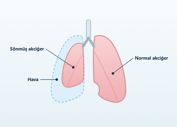
Pneumothorax is the partial or complete collapse of a lung caused by a buildup of air in the pleural space between the lung and the chest wall. It is also commonly known as a "collapsed lung."
Under normal conditions, there is no air in the pleural space. Air entering this space causes the lung to pull away from the chest wall and disrupts its normal expansion.
The impact of a pneumothorax does not depend only on how large it appears on imaging. In addition to the amount of air that has accumulated and how quickly it formed, the following also matter:
- The degree of shortness of breath
- Oxygenation
- Circulatory status
- Underlying lung disease
- The person's existing respiratory capacity
How Does a Pneumothorax Develop?
The outer surface of the lung is covered by a thin membrane called the visceral pleura, and the inner surface of the chest cavity by a thin membrane called the parietal pleura.
The pleural space between these two membranes helps the lung move in harmony with the chest wall during breathing.
Air entering the pleural space from the surface of the lung or from outside the chest wall disrupts this system. Air that builds up in the pleural space can cause the lung to pull away from the chest wall and partially or completely collapse.
If the air leak is small and closes on its own, the air remaining in the pleural space can be gradually absorbed by the body.
If the leak continues:
- The pneumothorax may enlarge
- The lung may not expand adequately
- The air leak may persist
What Are the Types of Pneumothorax?
Pneumothorax is classified into different groups based on its cause and the person's existing lung condition.
Primary Spontaneous Pneumothorax
Primary spontaneous pneumothorax is a pneumothorax that develops on its own, without significant trauma or previously known lung disease.This definition does not mean that there are no structural changes in the lung tissue. In some people, computed tomography may reveal blebs, bullae, or emphysema-like changes, particularly in the upper parts of the lung.
Although primary spontaneous pneumothorax is more common in young adults, it can also develop in people of different ages and body types. Smoking is one of the important risk factors. Not every bleb or bulla leads to a pneumothorax. In some people, these structures may be discovered incidentally on imaging performed for another reason.
Secondary Spontaneous Pneumothorax
Secondary spontaneous pneumothorax develops in people who have a previously known or clinically significant lung disease. Associated conditions may include:- COPD
- Emphysema
- Bullous lung disease
- Cystic fibrosis
- Bronchiectasis
- Pulmonary fibrosis and other interstitial lung diseases
- Tuberculosis
- Certain lung infections
- Lung cancer
- Lung metastases
- Lymphangioleiomyomatosis
- Certain connective tissue and genetic disorders
Traumatic Pneumothorax
Traumatic pneumothorax can develop after blunt or penetrating injuries to the chest. Possible causes include:- Traffic accidents
- Falls from a height
- Rib fractures
- Stab or penetrating wounds
- Blows to the chest
- Crush injuries
Pneumothorax Related to Medical Procedures
A pneumothorax can develop when the lung or pleura is affected during certain medical procedures. This is called iatrogenic pneumothorax. Procedures that may be associated with it include:- Lung needle biopsy
- Pleural biopsy
- Central venous catheter placement
- Thoracentesis
- Certain bronchoscopic procedures
- Mechanical ventilation
- Placement of certain cardiac devices
Catamenial Pneumothorax
Catamenial pneumothorax is a special type of spontaneous pneumothorax that is linked in timing to the menstrual period. It usually occurs in the days around the start of menstruation and is more common on the right side. It may be associated with thoracic endometriosis. Because it can tend to recur, evaluation and treatment may require joint assessment by different specialties, such as thoracic surgery and obstetrics and gynecology.Tension Pneumothorax
Tension pneumothorax is a potentially life-threatening condition in which air entering the pleural space cannot escape adequately, causing pressure inside the chest to rise progressively. The rising pressure can affect not only the affected lung but also:- The heart
- The great vessels
- The lung on the opposite side
- Circulation
What Are the Symptoms of Pneumothorax?
Symptoms may vary depending on the size of the pneumothorax, how quickly it develops, and the person's existing lung capacity.
Common symptoms include:
- Sudden chest pain
- Shortness of breath
- A stabbing sensation or pressure in the chest
- Rapid breathing
- Increased heart rate
- Cough
- Fatigue
- Reduced exercise capacity
- Severe shortness of breath
- Low oxygen levels
- Bluish discoloration (cyanosis)
- Low blood pressure
- Altered consciousness
- Circulatory failure
How Is Pneumothorax Diagnosed?
Diagnosis is based on evaluating the person's complaints, physical examination findings, and imaging results together.
Chest X-Ray
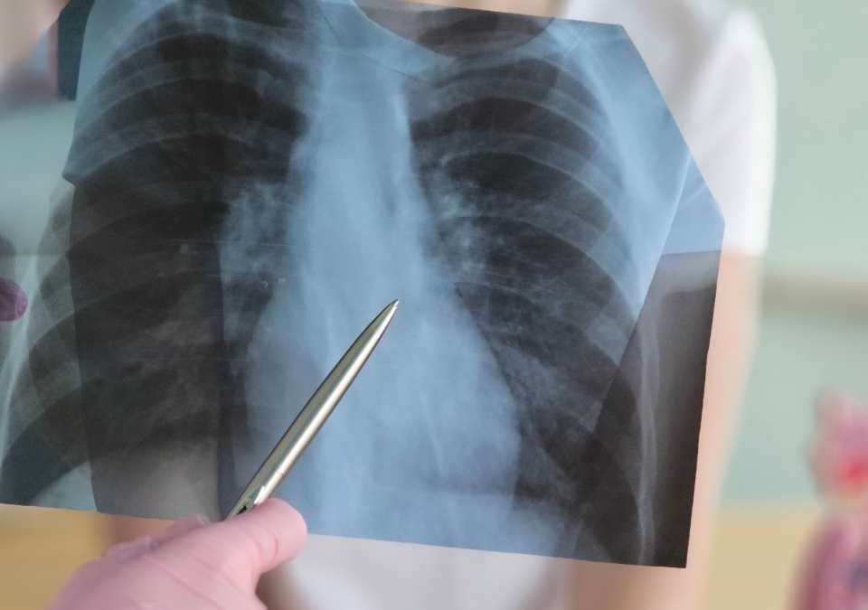
A chest X-ray is one of the basic imaging methods commonly used to diagnose pneumothorax. It can show the air in the pleural space and the degree of lung collapse. Very small pneumothoraces may not always be visible on a standard chest X-ray.Computed Tomography
Computed tomography shows the lungs and pleura in greater detail. Computed tomography can be used to evaluate: • Small pneumothoraces • Blebs or bullae • Areas of emphysema • Lung damage • Blood or fluid in the pleural space • Other trauma-related injuries • Underlying lung diseases Computed tomography is not necessary in every case of pneumothorax.Chest Ultrasound
Chest ultrasound can be used to evaluate pneumothorax, particularly in settings such as the emergency department and intensive care unit. An important advantage is that it can be performed at the bedside. The reliability of the result may depend on the operator's experience, the technique used, and the clinical conditions.Does the Size of a Pneumothorax Determine Treatment?
The size of a pneumothorax can be assessed with imaging, but in the current approach, treatment decisions are not based solely on a specific measurement in centimeters or on classifying the pneumothorax as "small" or "large."
The evaluation takes the following into account together:
- Chest pain
- The degree of shortness of breath
- Oxygenation
- Circulatory status
- The imaging characteristics of the pneumothorax
- Whether it is primary or secondary
- Underlying lung disease
- Whether it is a first or recurrent episode
- Whether it is on both sides
- Whether the air leak is continuing
- Whether safe follow-up is possible
How Is Pneumothorax Treated?
The goals of treatment are:
- Controlling the air in the pleural space
- Allowing the lung to re-expand
- Controlling symptoms
- Evaluating any ongoing air leak
- Reducing the risk of recurrence in suitable patients.
What Is a Part-Solid Nodule?
Conservative Observation
In some patients with primary spontaneous pneumothorax who are clinically stable and have no significant pain or shortness of breath, a conservative approach with close follow-up may be used. In this approach, the natural course of the pneumothorax is monitored instead of actively removing the air. If the air leak has stopped, the air in the pleural space can be gradually absorbed by the body. The decision to follow a pneumothorax conservatively is not based solely on its size on imaging. The availability of safe follow-up and the person's clinical characteristics are also important.Outpatient Treatment
In selected patients with primary spontaneous pneumothorax, outpatient treatment with special drainage systems that contain a one-way valve may be considered when an appropriate center and follow-up conditions are available. This approach is not suitable for every patient or every healthcare facility. Patient selection, close follow-up, and rapid access to the hospital when needed are important.Oxygen Therapy
Supplemental oxygen may be given to patients with low oxygen levels or who clinically need oxygen therapy. Not every patient with pneumothorax automatically needs high-flow oxygen. In some patients with chronic lung disease, especially those at risk of carbon dioxide retention, oxygen therapy is given in a controlled manner.Needle Aspiration
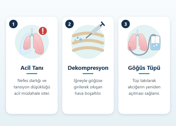
In needle aspiration, air in the pleural space is removed using a needle or a thin catheter. It may be one of the initial alternatives to a chest tube, particularly in some patients with primary spontaneous pneumothorax who require intervention. After the procedure, the re-expansion of the lung is evaluated. If air builds up again or adequate improvement is not achieved, a different drainage method may be needed.Chest Tube Placement
A chest tube is a drainage system placed between the ribs into the pleural space that allows the accumulated air to escape. After chest tube placement, the following are monitored:- Expansion of the lung
- Whether the air leak is continuing
- Respiratory signs
- The position of the tube
- Any accompanying fluid or blood in the pleural space.
How Is Tension Pneumothorax Treated?
Tension pneumothorax is a clinical emergency. The pressure inside the chest must be reduced quickly, followed by appropriate drainage of the pleural space. The treatment approach for tension pneumothorax differs from the routine management of a stable spontaneous pneumothorax.
When Is Surgical Treatment Considered for Pneumothorax?
Not every first-time spontaneous pneumothorax requires surgery.
The goal of surgery is to control an existing air leak and reduce the risk of recurrence in suitable patients.
Surgical evaluation may be considered particularly in the following situations:
- A second spontaneous pneumothorax on the same side
- A first pneumothorax on the opposite side
- Spontaneous pneumothorax on both sides
- Air leak continuing despite a chest tube
- The lung not re-expanding adequately
- Preventing recurrence being considered particularly important after a tension pneumothorax
- Spontaneous hemopneumothorax
- Catamenial pneumothorax
- An underlying disease that itself requires surgical treatment
- Recurrence posing a serious risk because of the person's occupation or living conditions
How Is Pneumothorax Surgery Performed?
Many pneumothorax operations can be performed using video-assisted thoracoscopic surgery (VATS).
During VATS, the chest cavity is accessed with a camera and surgical instruments through small incisions in the chest wall.
The extent of surgery may vary depending on the cause of the pneumothorax and the findings during the operation.
Bleb or Bulla Resection
Blebs or bullae thought to be causing the air leak can be removed together with a limited amount of surrounding lung tissue.
This is usually done using a surgical stapler.
If no obvious bleb or bulla is seen during surgery, this does not mean that the pneumothorax did not occur.
Some structural changes that cause air leaks may be very small or may not be clearly visible during surgery.
Pleural Procedures
Removing the bleb or bulla alone does not completely eliminate the risk of the pneumothorax recurring.
For this reason, procedures aimed at creating controlled adhesion between the lung and the chest wall may be performed during surgery.
These may include:
- Mechanical pleural abrasion
- Pleurectomy
- Chemical pleurodesis
Open Surgery
VATS is frequently used in pneumothorax surgery.
However, an open surgical approach may be considered in selected situations such as:
- Extensive pleural adhesions
- Complex lung disease
- An air leak that is difficult to control
- Bleeding
- Aiming for the lowest possible risk of recurrence in certain occupations
What Is a Persistent Air Leak?
When air continues to pass from the surface of the lung into the pleural space even after a chest tube has been placed, this is called a continuing or persistent air leak.
Some air leaks close quickly, while others may continue for days.
A persistent air leak may be associated with:
- Large or multiple bullae
- Emphysema
- Lung infection
- Pulmonary fibrosis
- Fragile lung tissue
- Trauma
- Previous lung surgery
- Inadequate lung expansion
- Autologous blood pleurodesis
- Chemical pleurodesis
- One-way drainage systems that can be used on an outpatient basis
- Endobronchial methods
Does Pneumothorax Recur?
Yes. Pneumothorax can recur after treatment.
The risk of recurrence varies depending on factors such as:
- The type of pneumothorax
- Underlying lung disease
- Smoking
- A previous pneumothorax
- The treatment given
- Smoking
- Blebs or bullae
- Emphysema
- Previous pneumothorax
- Family history
- Certain genetic disorders
- Thoracic endometriosis
- Chronic lung diseases
Is an Underlying Disease Investigated in Recurrent Pneumothorax?
Less common underlying diseases may be considered particularly in pneumothoraces that are:
- Recurrent
- Bilateral
- Familial
- Occurring at an unusual age
- Associated with atypical lung cysts on computed tomography
- Birt-Hogg-Dubé syndrome
- Marfan syndrome
- Certain Ehlers-Danlos syndromes
- Lymphangioleiomyomatosis
- Pulmonary Langerhans cell histiocytosis
- Thoracic endometriosis
- Alpha-1 antitrypsin deficiency
Recovery After Pneumothorax Surgery
After pneumothorax surgery, one or more chest tubes may be used to remove air and fluid from the pleural space.
Postoperative care may include:
- Pain control
- Breathing exercises
- Effective coughing
- Early mobilization
- Chest tube monitoring
- Lung imaging
- Monitoring for infection and bleeding
- Assessment of air leak
- The extent of the surgery performed
- Whether the air leak continues
- Underlying lung disease
- Overall health
Physical Activity After Pneumothorax
Return to physical activity is planned according to the complete resolution of the pneumothorax, the treatment given, and the person's clinical condition.
Light daily activities and walking can be continued as symptoms allow.
On the other hand, the following may be postponed until the pneumothorax has fully resolved:
- Intense exercise
- Heavy lifting
- Contact sports
- Excessive physical exertion
Air Travel After Pneumothorax
Pressure changes in an airplane cabin can cause air in the pleural space to expand.
For this reason, commercial air travel is not appropriate while a pneumothorax is still present.
Before flying, complete resolution of the pneumothorax must be confirmed with imaging.
Even after complete radiological resolution has been shown, an additional waiting period may be recommended because of the possibility of early recurrence. International guidelines include an approach of waiting at least 7 days after complete radiological resolution has been demonstrated.
However, the following may affect the assessment for flying:
- The cause of the pneumothorax
- The treatment given
- Recent surgery
- Underlying lung disease
- Airline- and country-specific rules
Diving After Pneumothorax
The significant pressure changes that occur during scuba diving pose a particular risk for people who have previously had a spontaneous pneumothorax.
Returning to scuba diving after a history of spontaneous pneumothorax is evaluated differently from returning to everyday exercise or air travel.
Because a recurrent pneumothorax developing underwater can have serious and potentially life-threatening consequences, scuba diving is generally not recommended after a spontaneous pneumothorax.
In certain special cases, people who have undergone definitive surgery to prevent recurrence may be evaluated separately from a diving medicine perspective. However, having had surgery does not by itself automatically mean that a person is fit to dive.
The pressure and gas dynamics of free diving and scuba diving are not the same. Even so, a history of pneumothorax should be taken into account when evaluating both activities.
Smoking and Pneumothorax
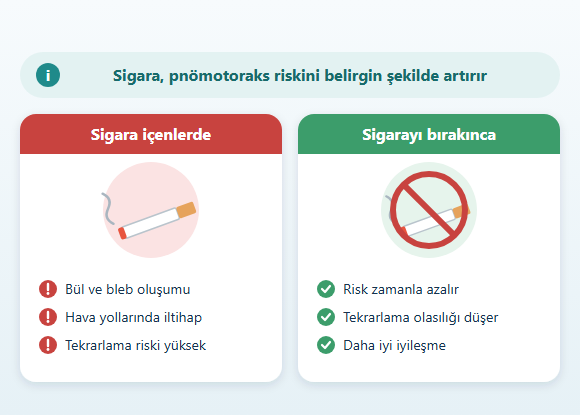
Smoking is associated with the development and recurrence of pneumothorax, particularly primary spontaneous pneumothorax.
Quitting smoking does not guarantee that a pneumothorax will not recur, but it is important for protecting lung health and reducing modifiable risk factors.
E-cigarettes, hookah (water pipe), and other inhaled products should not be considered risk-free for the lungs either.
Especially in people who have previously had a pneumothorax, current lung health should be taken into account when considering the use of inhaled substances.
Pneumothorax and Family History
Pneumothorax can occur in more than one member of some families. A family history of:- Recurrent pneumothorax
- Kidney tumors
- Certain skin lesions
- Connective tissue disease
- Cystic lung disease
Follow-Up After Pneumothorax
The follow-up plan is determined by:
- The type of pneumothorax
- The treatment given
- Underlying lung disease
- The risk of recurrence
- The course of symptoms
- Re-expansion of the lung
- Resolution of the air leak
- The chest tube site or surgical wound
- Underlying lung disease
- Factors that may increase the risk of recurrence
Frequently Asked Questions About Pneumothorax
Is pneumothorax the same as a collapsed lung?
Pneumothorax is commonly referred to as a "collapsed lung." A buildup of air in the pleural space causes the lung to pull away from the chest wall and partially or completely collapse.
Can a pneumothorax go away on its own?
Some primary spontaneous pneumothoraces in patients who are clinically stable and have few symptoms can be observed without intervention and may resolve over time. However, this approach is not suitable for every patient with pneumothorax.
Does a large pneumothorax always require a chest tube?
No. In the current approach, particularly in primary spontaneous pneumothorax, treatment is not determined solely by the size seen on imaging. Symptoms, physiological status, underlying lung disease, and the availability of safe follow-up are evaluated together.
Why does pneumothorax recur?
Blebs or bullae in the lung, smoking, emphysema, certain chronic or genetic lung diseases, and a previous pneumothorax may be associated with the risk of recurrence.
Is surgery needed for a first pneumothorax?
In most patients, a first spontaneous pneumothorax does not automatically require surgery. However, surgery may be considered after a first episode in certain situations, such as tension pneumothorax, high-risk occupations, or when preventing recurrence is particularly important.
What is done during pneumothorax surgery?
During surgery, blebs or bullae that may be causing an air leak can be removed, and pleural procedures can be performed to reduce the risk of the pneumothorax recurring. Surgery can usually be performed using VATS.
What happens if the air leak through the chest tube continues?
The duration and volume of the air leak and the expansion of the lung are evaluated. A prolonged air leak may require surgical evaluation. In some patients who are not suitable for surgery, other pleural or bronchoscopic methods may be used.
Can I fly after having a pneumothorax?
Flying is not recommended while a pneumothorax is present. First, it must be confirmed with imaging that the lung has fully re-expanded. A certain waiting period may also be needed after complete radiological resolution.
Can a person who has had a pneumothorax go scuba diving?
A history of spontaneous pneumothorax poses a significant risk for scuba diving, and diving is generally not recommended. Even after definitive surgery to prevent recurrence, fitness to dive requires a separate, specialized evaluation.
Does everyone who has a pneumothorax need a CT scan?
No. Computed tomography is not required in every case of pneumothorax. It may be used for a more detailed evaluation in situations such as underlying lung disease, recurrent pneumothorax, trauma, diagnostic uncertainty, or surgical planning.
Disclaimer: The information on this website is intended for general informational purposes only and is not a substitute for a personal medical evaluation. Decisions about the diagnosis, follow-up, and treatment of pneumothorax are made individually by the physician based on the person's symptoms, clinical condition, imaging findings, underlying lung diseases, and overall health.