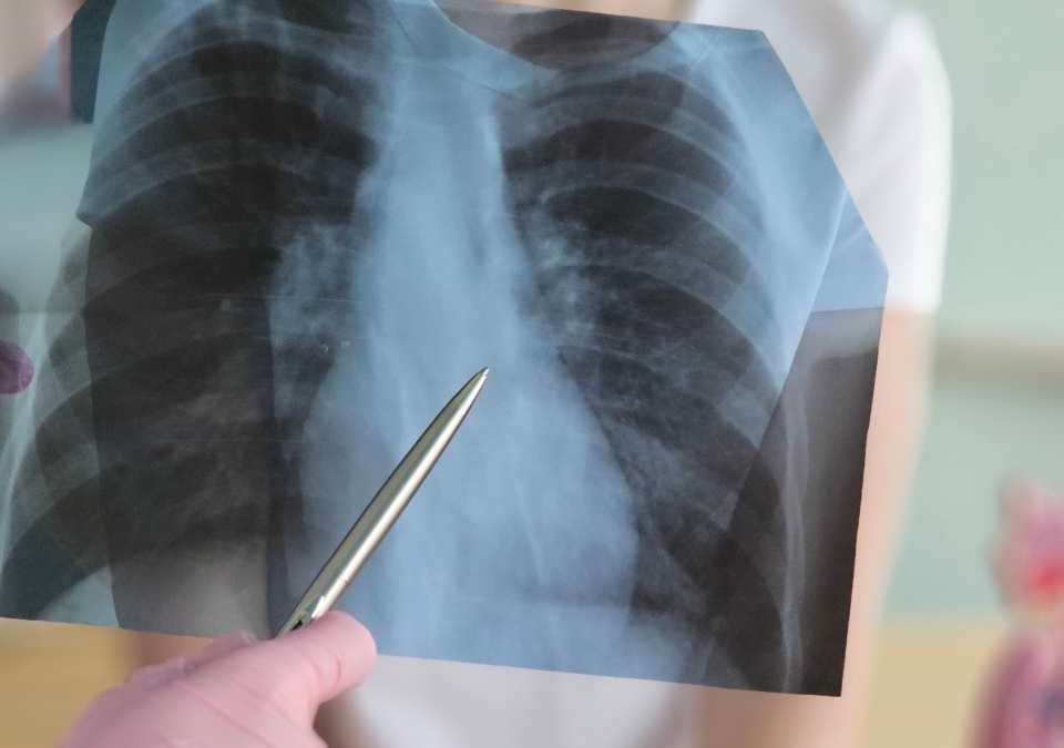
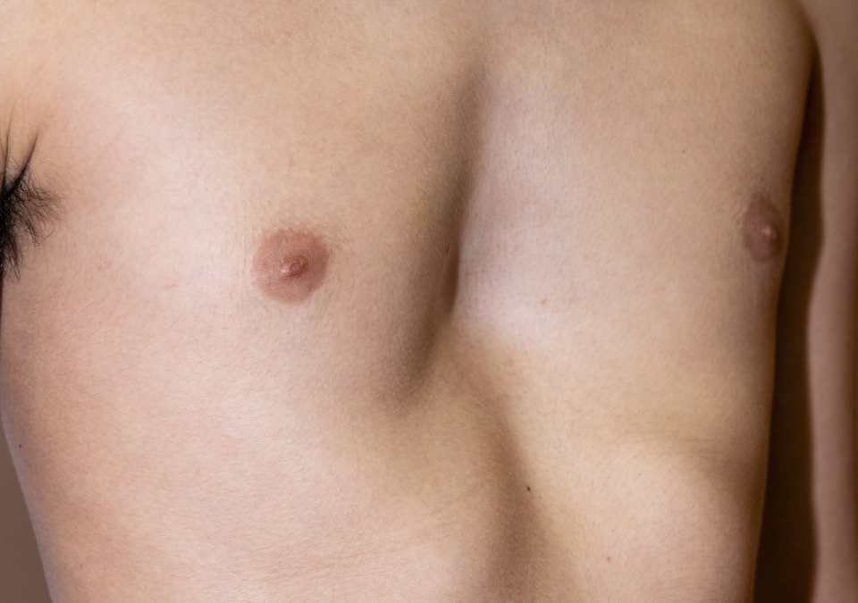
Mediastinal and Thymic Diseases

Information on Mediastinal Masses, Thymoma, Thymic Diseases, the Link with Myasthenia Gravis, Diagnosis, Biopsy, Staging, and Surgical Treatment Methods.
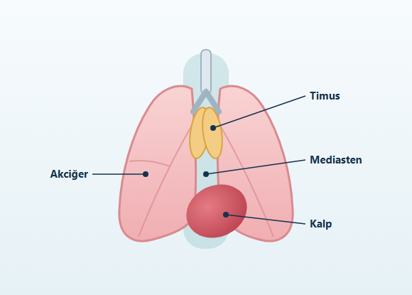
What Are Mediastinal and Thymic Diseases?

The mediastinum is the anatomical region of the chest cavity located between the two lungs. This area contains the heart, the great vessels, the windpipe (trachea), the main bronchi, the esophagus, lymph nodes, nerve structures, and the thymus gland. Diseases of the mediastinum do not belong to a single organ. Benign or malignant tumors, cysts, enlarged lymph nodes, nerve-derived growths, thyroid tissue, and thymic diseases can all occur in this region. Some mediastinal lesions are found incidentally on imaging performed for another reason and cause no symptoms. Others may press on surrounding organs and tissues, causing complaints such as cough, shortness of breath, chest pain, difficulty swallowing, or hoarseness. The diagnostic and treatment plan is determined by the lesion's location within the mediastinum, its imaging features, its size, its relationship to surrounding tissues, the likely type of disease, and the person's overall health. Not every mediastinal mass requires surgery, and not every mediastinal mass needs a biopsy before surgery.

What Are the Compartments of the Mediastinum?

The mediastinum is divided into compartments to describe the location of lesions detected on imaging and to classify possible diseases. In the current computed tomography-based classification, the mediastinum is generally considered to consist of:
  • The anterior or prevascular compartment
  • The middle or visceral compartment
  • The posterior or paravertebral compartment
The compartment in which a mass is located does not by itself establish a definitive diagnosis. However, it can help narrow down the possible diseases, determine which imaging methods are needed, and plan the biopsy or surgical approach.

Anterior Mediastinum or Prevascular Compartment

The anterior mediastinum is the region behind the breastbone and in front of the heart and great vessels. Lesions that may be found in this area include:
  • Thymoma
  • Thymic carcinoma
  • Thymic cysts
  • Thymic hyperplasia
  • Lymphoma
  • Germ cell tumors
  • Thyroid tissue extending into the mediastinum
  • Parathyroid-derived lesions
  • Certain tumors arising from fatty tissue
Thymic diseases are most often found in this compartment.

Middle Mediastinum or Visceral Compartment

The visceral mediastinum is the anatomical region associated with the trachea, main bronchi, esophagus, heart, and great vessels. The following may be seen in this region:
  • Enlarged lymph nodes
  • Bronchogenic cysts
  • Pericardial cysts
  • Esophageal lesions
  • Vascular dilations and vascular lesions
  • Diseases related to the trachea and bronchi
It should be kept in mind that some structures that appear as a mass on imaging may be vascular in origin. For this reason, the relationship of the lesion to the great vessels is evaluated in detail, especially before a biopsy or surgical procedure.

Posterior Mediastinum or Paravertebral Compartment

The paravertebral mediastinum is the region close to the spine. A significant proportion of masses found in this region may originate from nerves or nerve sheath tissue. Lesions that may be seen include:
  • Schwannoma
  • Neurofibroma
  • Ganglioneuroma
  • Other neurogenic tumors
  • Spine-related masses
  • Certain cysts of esophageal origin
  • Paravertebral infections
Some neurogenic tumors may extend into the spinal canal. When this is a possibility, magnetic resonance imaging is particularly important for evaluating the relationship with the spinal cord and nerve structures.

What Are the Symptoms of Mediastinal Diseases?

Mediastinal lesions may cause no symptoms for a long time. Small masses that do not press on surrounding structures may be noticed incidentally on a chest X-ray or computed tomography scan taken for another reason. When symptoms do occur, they may vary depending on the mass's location, its size, and the anatomical structures it affects. Possible symptoms include:
  • Chest pain or a feeling of pressure in the chest
  • Shortness of breath
  • Cough
  • Hoarseness
  • Difficulty swallowing
  • Back or shoulder pain
  • Swelling of the face and neck
  • Prominent neck veins
  • Recurrent lung infections
  • Fever
  • Night sweats
  • Fatigue
  • Loss of appetite
  • Unintentional weight loss
Compression of the great vessels can lead to swelling of the face, neck, and arms. Involvement of the trachea can cause shortness of breath or abnormal sounds during breathing. Pressure on the esophagus can cause difficulty swallowing. Involvement of the nerves responsible for vocal cord movement can lead to hoarseness. General symptoms such as fever, night sweats, and unintentional weight loss may be seen in lymphoma, infections, or certain malignant diseases. However, these symptoms are not specific to any particular mediastinal disease.

What Is a Mediastinal Mass?

A mediastinal mass is a general term for an abnormal lesion detected on imaging in the region between the two lungs. This term does not mean that the mass is cancer. A mass-like appearance in the mediastinum can be caused by very different conditions, such as:
  • Tumors
  • Cysts
  • Enlarged lymph nodes
  • Vascular structures
  • Infections
  • Congenital developmental lesions
  • Thyroid or parathyroid tissue
When evaluating a mediastinal mass, features such as the following are taken into account:
  • Its location in the mediastinum
  • Size
  • Margin characteristics
  • Whether it is solid, fluid-filled, or mixed
  • Whether it contains fat or calcium
  • Its relationship to the great vessels
  • Extension into the lung, pleura, or pericardium
  • Growth or change compared with previous imaging

How Are Mediastinal Masses Evaluated?

Imaging plays a central role in the evaluation of mediastinal masses. Depending on the suspected disease, blood tests, neurological examinations, endocrinological evaluations, and tissue sampling may also be needed.

Chest X-Ray

A chest X-ray may be the first test in which mediastinal widening or a mass is noticed. However, because it provides a two-dimensional image, it gives limited information about the exact location and structure of the mass. For this reason, computed tomography is usually used for detailed evaluation.

Computed Tomography

Contrast-enhanced chest computed tomography is one of the main imaging methods for evaluating mediastinal masses. Computed tomography can be used to assess:
  • Location of the mass
  • Size
  • Internal structure
  • Cystic or solid features
  • Relationship to the great vessels
  • Lung or pleural involvement
  • Status of the lymph nodes
  • Extension to the chest wall or pericardium
  • Calcification
  • Fat content
The use of intravenous contrast can be especially important in distinguishing a vascular structure from a tumor.

Magnetic Resonance Imaging

Magnetic resonance imaging allows detailed evaluation of soft tissues and certain anatomical relationships. It may be used particularly for:
  • Evaluating cystic lesions
  • Distinguishing thymic hyperplasia from certain thymic masses
  • Paravertebral masses that may extend into the spinal canal
  • Lesions closely related to the heart and great vessels
  • Tissue characterization in selected cases
Magnetic resonance imaging is not necessary for every mediastinal mass.

PET-CT

PET-CT is an imaging method that evaluates the metabolic activity of tissues. Particularly in lymphoma, thymic tumors, and certain other malignant diseases, it may be used for:
  • Assessing the extent of the disease
  • Examining the lymph nodes
  • Looking for distant organ involvement
  • Identifying a suitable biopsy site
  • Evaluating treatment response in certain diseases
Infections and inflammatory diseases can also show high metabolic activity on PET-CT. For this reason, increased uptake on PET-CT does not by itself mean a cancer diagnosis.

Blood Tests and Tumor Markers

Depending on the location of the mediastinal mass and the type of disease suspected, certain blood tests can help in the diagnostic process. Especially when a germ cell tumor is possible in the anterior mediastinum, values such as the following may be checked:
  • Alpha-fetoprotein (AFP)
  • Beta human chorionic gonadotropin (beta-hCG)
  • Lactate dehydrogenase (LDH)
When a thyroid-derived lesion is suspected, thyroid function tests may be used. In people who need evaluation for myasthenia gravis, acetylcholine receptor antibodies and, depending on the clinical situation, other immune tests may be used. Blood tests alone do not establish a definitive diagnosis of a mediastinal mass. The findings are evaluated together with imaging and, when necessary, pathology results.

Is Every Mediastinal Mass Biopsied?

No. Not every mediastinal mass needs a biopsy before surgery. The decision to biopsy is made by taking into account:
  • The imaging features of the mass
  • The likely type of disease
  • Whether it appears surgically completely removable
  • Whether the biopsy result would change the treatment plan
  • Its relationship to the great vessels and heart
  • Bleeding risk
  • The safety of the chosen biopsy route
  • The person's overall health
If lymphoma, a germ cell tumor, or another disease whose first-line treatment may not be surgery is suspected, obtaining an adequate tissue sample is important. On the other hand, a preoperative biopsy may not be necessary for some anterior mediastinal masses whose imaging features are consistent with a thymic epithelial tumor and which appear well-circumscribed and surgically completely removable. When a biopsy is needed, it is important to choose a method that will not adversely affect a possible surgical plan. Methods that may be used include:
  • Computed tomography-guided needle biopsy
  • Ultrasound-guided biopsy
  • Bronchoscopy
  • Endobronchial ultrasound (EBUS)
  • Endoscopic ultrasound
  • Mediastinoscopy
  • Mediastinotomy
  • VATS biopsy
  • Biopsy of accessible peripheral lymph nodes
The method is determined by the location of the mass, the suspected disease, and a safe access route.

What Is Mediastinoscopy?

Mediastinoscopy is a procedure that allows access to the mediastinum with a camera and surgical instruments through a small surgical incision near the lower part of the neck.

It can be used particularly to obtain tissue samples from certain lymph nodes around the trachea or from suitably located mediastinal lesions. Today, many mediastinal lymph nodes can be sampled with ultrasound-assisted bronchoscopic methods such as EBUS.

However, in some cases where these methods do not yield an adequate sample or a diagnosis, surgical biopsy methods may be needed. Mediastinoscopy is not suitable for every mediastinal mass. The decision to perform the procedure is based on the location of the mass and the suspected disease.

What Is the Thymus Gland?

The thymus is an organ related to the immune system, located behind the breastbone and in front of the heart. It plays an important role in the development of the immune system during childhood. It contributes to the maturation of immune cells called T lymphocytes. During adolescence and adulthood, much of the thymic tissue shrinks and is partly replaced by fatty tissue. However, the thymus does not disappear completely in adults. Diseases that may occur in the thymus include:
  • Thymic hyperplasia
  • Thymic cysts
  • Thymoma
  • Thymic carcinoma
  • Thymic neuroendocrine tumors
  • Fat-related lesions such as thymolipoma

Thymic Hyperplasia

Thymic hyperplasia is a condition in which the thymic tissue appears larger than normal or more active at the cellular level. It may be associated with myasthenia gravis and certain immune system diseases. Regrowth of thymic tissue that has shrunk after chemotherapy, severe infection, or major physical stress can also create the appearance of hyperplasia on imaging. Thymic hyperplasia is not a thymic tumor. However, in some cases the imaging findings may be confused with thymic masses. Computed tomography and, in selected cases, magnetic resonance imaging can help make the distinction.

Thymic Cyst

Thymic cysts are fluid-filled lesions that can arise in thymic tissue or along the embryological developmental path of the thymus. Thymic cysts may be congenital or acquired. Some are simple, thin-walled, and single-chambered, while others may have more complex features. Thymic cysts usually cause no symptoms. Large cysts may press on surrounding structures and cause complaints such as:
  • Chest pain
  • Cough
  • Shortness of breath
The decision between observation and surgery is based on factors such as:
  • Size of the cyst
  • Imaging features
  • Whether it causes symptoms
  • Growth over time
  • Diagnostic uncertainty

What Is Thymoma?

Thymoma is one of the rare tumors that develop from the epithelial cells of the thymus gland and is usually found in the anterior mediastinum. Thymomas can show different biological behaviors. Some tumors may be confined and slow-growing, while others may extend into the surrounding:
  • Mediastinal fat
  • Pleura
  • Lung
  • Pericardium
  • Great vessels
Thymoma does not always cause symptoms. In some patients, it may be found incidentally during imaging performed for another reason. When symptoms are present, they may include:
  • Chest pain
  • Cough
  • Shortness of breath
  • A feeling of pressure in the chest
  • Difficulty swallowing
  • Swelling of the face or neck
One of the important features of thymoma is that it can occur together with certain autoimmune and neurological diseases, the foremost of which is myasthenia gravis.

What Is Thymic Carcinoma?

Thymic carcinoma also develops from thymic epithelial cells, but it has biological and pathological features that differ from thymoma. Compared with thymoma, thymic carcinomas may generally follow a different course in terms of:
  • More invasive behavior toward surrounding tissues
  • Spread to lymph nodes
  • Metastasis to the pleura, lung, or distant organs
The distinction between thymoma and thymic carcinoma can be suggested by imaging, but the definitive diagnosis is made by pathological examination. Depending on the stage of the disease and whether it can be removed, treatment may include different combinations of surgery, radiation therapy, and systemic therapies.

Thymic Neuroendocrine Tumors

Thymic neuroendocrine tumors are rare tumors that develop from the thymus. They have pathological and biological features that differ from thymoma and thymic carcinoma. Some may be associated with hormone production or certain hereditary endocrine diseases. The treatment plan is determined by taking into account:
  • Tumor size
  • Spread to surrounding tissues
  • Status of the lymph nodes
  • Distant organ metastasis
  • Hormonal activity
Surgery may be part of treatment for completely removable tumors. In advanced disease, systemic therapies or radiation therapy may be considered.

What Is the Relationship Between Myasthenia Gravis and the Thymus?

Myasthenia gravis is an autoimmune disease that causes fluctuating weakness in voluntary muscles as a result of impaired transmission from nerves to muscles. The thymus gland may play a role in the immune-related mechanisms of myasthenia gravis. Thymic hyperplasia or thymoma may be found in some people with myasthenia gravis. Symptoms may include:
  • Drooping eyelids
  • Double vision
  • Weakness of the facial muscles
  • Fatigue while chewing
  • Difficulty swallowing
  • Impaired speech
  • Weakness of the neck, arm, or leg muscles
  • Muscle fatigue that may worsen over the course of the day
  • Weakness of the respiratory muscles
A person diagnosed with thymoma may undergo clinical evaluation for myasthenia gravis even if they have no obvious muscle complaints. The diagnostic process may include:
  • Neurological examination
  • Antibody tests
  • Nerve conduction studies
  • Electromyography
  • Chest imaging
Because involvement of the respiratory or swallowing muscles can lead to more serious clinical consequences, preoperative neurological evaluation is particularly important in patients with myasthenia gravis.

Thymus Surgery and Myasthenia Gravis

Surgical removal of the thymus is called thymectomy. In people with thymoma, removal of the thymoma and the thymic tissue is considered when surgically appropriate. The presence of myasthenia gravis requires separate planning of management before and after surgery. In some patients with generalized myasthenia gravis who do not have a thymoma, thymectomy may also be part of the neurological treatment plan, particularly when the type of disease and antibody profile are suitable. The effect of thymectomy on myasthenia gravis does not necessarily appear immediately after surgery. A reduction in muscle weakness or a change in medication needs may develop over months or longer. Having had a thymectomy does not mean that myasthenia gravis will definitely go away. Neurological follow-up and any necessary medications may continue after surgery.

How Is Thymoma Staged?

Staging of thymic epithelial tumors is based on a combined assessment of features such as tumor size, invasion into surrounding structures, lymph nodes, and distant spread. In the current TNM system, the evaluation takes into account features such as:
  • Tumor size
  • Relationship to the mediastinal fat
  • Pleural involvement
  • Pericardial involvement
  • Lung invasion
  • Involvement of the great vessels or other mediastinal structures
  • Lymph node spread
  • Pleural or pericardial spread
  • Distant organ metastasis
Before surgery, the clinical stage can be estimated using imaging methods. In patients who undergo surgery, the pathological stage can be determined from pathological examination of the removed tumor and related tissues. Whether the tumor was completely removed and the status of the surgical margins are also important for subsequent treatment and follow-up planning.

How Is Thymoma Treated?

Thymoma treatment is planned by taking into account:
  • Stage of the disease
  • Whether the tumor can be completely removed
  • Its relationship to surrounding tissues
  • Whether myasthenia gravis is present
  • The person's overall health
For confined and completely removable thymomas, surgery is one of the main treatment options. The goal of surgery is to remove the tumor and the necessary thymic tissue with appropriate surgical margins. During the operation, it is important that the tumor is not fragmented and that tumor tissue does not spread into the chest cavity. For some tumors that have extended into neighboring organs or initially do not appear completely removable, the following may be used in different sequences or combinations:
  • Systemic therapy
  • Surgery
  • Radiation therapy
Because thymic tumors are rare, multidisciplinary evaluation is important in treatment planning.

How Is Thymus Surgery Performed?

The method used in thymus surgery is determined by the tumor's size, location, relationship to surrounding tissues, and the extent of the surgical procedure required.

VATS Thymus Surgery

Video-assisted thoracoscopic surgery, or VATS, is a minimally invasive method performed through small incisions using a camera and surgical instruments. It may be used for some thymic diseases that do not show significant invasion into surrounding structures and are thought to be safely removable in one piece with a minimally invasive approach. The main goal of minimally invasive surgery is not simply to make small incisions. If oncologic surgery is being performed, the tumor must be removed with appropriate margins while keeping the tumor intact.

Robot-Assisted Thymus Surgery

Robot-assisted surgery is also one of the minimally invasive methods performed through small incisions. The robotic system can provide a magnified three-dimensional view and greater range of motion within the narrow space of the mediastinum. However, robot-assisted surgery is not suitable for every thymic tumor. The tumor's relationship to the following is taken into account when choosing the surgical approach:
  • Great vessels
  • Lung
  • Pericardium
  • Trachea
  • Other mediastinal structures

Sternotomy

Sternotomy is an open surgical method in which the mediastinum is accessed by dividing the breastbone along the midline. In large, centrally located, or locally advanced thymic tumors, it can provide wide surgical exposure and access. Sternotomy or more extensive open approaches may be used when additional surgical procedures are needed on the great vessels, pericardium, or other mediastinal structures.

Thoracotomy

Thoracotomy is an open surgical method in which the chest cavity is accessed between the ribs. It may be used in selected cases where the tumor extends predominantly into one side of the chest cavity, the lung, or the pleura. The key criterion in choosing the surgical method is that the required procedure can be performed safely and in an oncologically appropriate manner.

What Tests Are Performed Before Thymus Surgery?

Before surgery, both the extent of the tumor and the person's fitness for surgery are evaluated. When necessary, the following may be performed:
  • Contrast-enhanced chest computed tomography
  • Magnetic resonance imaging
  • PET-CT
  • Pulmonary function tests
  • Cardiac evaluation
  • Blood tests
  • Antibody tests for myasthenia gravis
  • Neurological evaluation
  • Anesthesia evaluation
  • Biopsy
In people with myasthenia gravis, muscle strength, swallowing function, respiratory muscles, and current medications are evaluated separately. In some cases where the disease is not adequately controlled, the neurological treatment may need to be adjusted before surgery.

Recovery After Thymus Surgery

The postoperative course varies according to the surgical method, the extent of tissue removed, and the person's overall health. After surgery, the following may be needed:
  • Pain control
  • Breathing exercises
  • Early mobilization
  • Chest tube monitoring
  • Monitoring for bleeding and infection
  • Assessment of muscle strength and swallowing function
  • Adjustment of myasthenia gravis treatment
In people with myasthenia gravis, closer monitoring may be needed because of the risk of respiratory muscle weakness. The removed tissue is examined in the pathology laboratory. The pathology report may assess:
  • The exact type of tumor
  • Histological features
  • Tumor size
  • Spread to surrounding tissues
  • Surgical margins
  • Lymph node status
  • Pathological stage
Whether additional radiation therapy or systemic therapy is needed after surgery is determined according to the pathology result and the characteristics of the disease.

Mediastinal Cysts

Mediastinal cysts may be congenital developmental structures or acquired cystic lesions. The main types of cysts include:
  • Bronchogenic cyst
  • Pericardial cyst
  • Thymic cyst
  • Esophageal cysts
  • Neurenteric cysts
  • Lymphatic cysts
Cysts may cause no symptoms for a long time. In case of growth, infection, bleeding, or pressure on surrounding organs, the following may occur:
  • Cough
  • Shortness of breath
  • Chest pain
  • Difficulty swallowing
  • Recurrent infections
Some cysts that have typical imaging features and cause no symptoms can be monitored. Surgical removal may be considered for cysts that cannot be definitively diagnosed, are growing, cause symptoms, or carry a risk of complications.

Bronchogenic Cyst

Bronchogenic cysts are among the congenital structures that form during the development of the respiratory system. They are usually located near the trachea and main bronchi. Large bronchogenic cysts may press on:
  • The airways
  • The esophagus
  • Other surrounding mediastinal structures
The cysts may become infected or may be confused with other mediastinal masses on imaging. If surgery is planned, the cyst's relationship to the airways, esophagus, and great vessels is evaluated.

Pericardial Cyst

A pericardial cyst is a benign, usually fluid-filled lesion associated with the membrane surrounding the heart. It is often found incidentally on imaging performed for another reason. Some pericardial cysts that cause no symptoms and have typical imaging features can be monitored. In case of growth, symptoms, or diagnostic uncertainty, surgical evaluation may be needed.

Lymphoma and the Mediastinum

Lymphoma is a group of diseases that develop from cells of the lymphatic system. Some types of lymphoma can form large masses or enlarged lymph nodes, particularly in the anterior mediastinum. Possible symptoms include:
  • Lymph node enlargement
  • Fever
  • Night sweats
  • Unintentional weight loss
  • Itching
  • Fatigue
  • Cough
  • Shortness of breath
  • Swelling of the face and neck
The main treatment for lymphoma is usually not surgical removal of the entire mass. The main goal in diagnosis is to obtain a tissue sample of sufficient quantity and quality to determine the exact type of the disease. For this reason, the biopsy method is chosen carefully. In some cases, fine-needle aspiration alone may not be sufficient to determine the lymphoma subtype.

Mediastinal Germ Cell Tumors

Although germ cell tumors are most often found in the gonads, they can also develop in the mediastinum. In addition to benign lesions such as mature teratoma, there may be malignant germ cell tumors with different biological characteristics. The evaluation may use:
  • Computed tomography
  • PET-CT when necessary
  • AFP
  • Beta-hCG
  • LDH
  • Tissue sampling when needed
Treatment varies considerably depending on the type of tumor. While surgery may be the main treatment for mature teratomas, systemic drug therapies may take priority in some malignant germ cell tumors. For this reason, when a germ cell tumor is possible in an anterior mediastinal mass, evaluation of tumor markers is important for the biopsy and treatment plan.

Neurogenic Mediastinal Tumors

Neurogenic tumors develop from nerves or nerve sheaths and are mostly found in the paravertebral mediastinum. In adults, a significant proportion of these lesions may be benign, but malignant neurogenic tumors can also occur. Small masses may cause no symptoms. As the lesions grow, the following may appear:
  • Back pain
  • Chest pain
  • Numbness
  • Muscle weakness
  • Signs due to nerve root or spinal cord compression
When extension into the spinal canal is possible, magnetic resonance imaging is particularly important. Surgical treatment is planned by taking into account the size of the tumor, its relationship to the nerves, and its extension into the spinal canal. In some cases, a joint evaluation by thoracic surgery and neurosurgery may be needed.

Mediastinal Goiter

Extension of the thyroid gland into the chest cavity may be called mediastinal or retrosternal goiter. Large mediastinal goiters can cause:
  • Compression of the trachea
  • Shortness of breath
  • Cough
  • Difficulty swallowing
  • Fullness in the neck
  • Hoarseness
Computed tomography helps evaluate the extent of thyroid tissue in the mediastinum and its relationship to the great vessels and airways. The surgical approach is determined by the degree to which the thyroid tissue extends into the mediastinum and its relationship to surrounding structures.

Surgical Treatment of Mediastinal Diseases

Surgical treatment is not performed for the same purpose in every mediastinal disease. The goals of surgery may include:
  • Obtaining sufficient tissue for a definitive diagnosis
  • Removing a benign but growing mass
  • Relieving symptoms caused by compression
  • Treating or preventing complications of cysts
  • Removing a malignant tumor with appropriate surgical margins
  • Evaluating the lymph nodes when necessary
Surgical approaches that may be used include:
  • VATS
  • Robot-assisted surgery
  • Sternotomy
  • Thoracotomy
  • Neck incision
  • A combination of more than one approach in selected cases
Minimally invasive surgery is not suitable for every mediastinal mass. The tumor's relationship to the following is important in choosing the surgical method:
  • Great vessels
  • Heart
  • Trachea
  • Lung
  • Pleura
  • Nerve structures

A Multidisciplinary Approach to Mediastinal Diseases

Mediastinal diseases can originate from different structures such as the thymus, the lymphatic system, nerve tissue, the thyroid, the esophagus, and blood vessels. For this reason, depending on the characteristics of the disease, the evaluation may involve different specialties such as:
  • Thoracic Surgery
  • Pulmonology
  • Medical Oncology
  • Radiation Oncology
  • Radiology
  • Pathology
  • Neurology
  • Hematology
  • Endocrinology
  • Neurosurgery
  • Cardiovascular Surgery
Particularly in thymic tumors, lymphomas, germ cell tumors, and mediastinal masses extending into the great vessels, the sequence of treatments may be determined through multidisciplinary evaluation.

Follow-Up After Treatment

The follow-up program is determined by the type of disease, the pathology result, and the treatment received. Follow-up may include:
  • Clinical evaluation
  • Computed tomography
  • Magnetic resonance imaging or PET-CT when necessary
  • Assessment of neurological symptoms
  • Monitoring of pulmonary function
  • Evaluation of any additional treatments given
  • Checking for recurrence of the disease
For thymic epithelial tumors, follow-up may be long-term depending on the biological characteristics of the disease. Because thymomas in particular can sometimes recur long after the initial treatment, follow-up may not be limited to the first few years after surgery. If the disease recurs, the treatment plan does not automatically consist of repeating the previous treatment. The following aspects of the recurrence are re-evaluated:
  • Location
  • Extent
  • Previous treatments
  • Surgical resectability
  • The person's overall health

Frequently Asked Questions About Mediastinal and Thymic Diseases

Does a mediastinal mass mean cancer?
No. A mediastinal mass simply describes a lesion detected on imaging. Cysts, benign tumors, enlarged lymph nodes, vascular structures, thyroid tissue, and malignant tumors can all appear as a mass in the mediastinum.
Is a mediastinal mass always biopsied?
No. The decision to biopsy is based on the likely disease and the imaging features of the mass. While a biopsy is important in diseases such as lymphoma, where the tissue diagnosis determines treatment, some thymic tumors that appear completely removable may proceed directly to surgical evaluation.
Is thymoma cancer?
Thymoma is a thymic epithelial tumor that develops from the epithelial cells of the thymus. Its biological behavior can vary from patient to patient. Some thymomas remain confined, while others may extend into surrounding tissues or spread to the pleura.
Are thymoma and thymic carcinoma the same disease?
No. Although both can develop from thymic epithelial cells, their pathological and biological behavior differs. Thymic carcinoma can generally show more aggressive features.
Is there a link between thymoma and myasthenia gravis?
Yes. Myasthenia gravis may occur in some people with thymoma. For this reason, patients diagnosed with thymoma may be clinically evaluated for myasthenia gravis even if they have no obvious muscle complaints.
Does everyone with myasthenia gravis have thymus surgery?
No. The decision to perform a thymectomy is based on the type of myasthenia gravis, the patient's age, antibody profile, the extent of the disease, and whether a thymoma is present. In patients with thymoma, the surgical approach is also planned according to the characteristics of the tumor.
Can thymus surgery be performed with a minimally invasive (closed) technique?
In suitable patients, VATS or robot-assisted minimally invasive methods may be used. However, the size of the tumor and its relationship to the surrounding great vessels, lung, pericardium, or other organs may require open surgery.
Should mediastinal cysts be operated on?
Not every mediastinal cyst needs surgery. The type of cyst, its size, imaging features, whether it is growing, and whether it causes symptoms or complications all influence the decision.
Can thymoma recur after surgery?
Yes. The risk of recurrence depends on factors such as the stage of the tumor, its pathological features, and whether it was completely removed surgically. Because some thymomas can recur long afterward, long-term follow-up may be needed.
Which specialty should evaluate a mediastinal mass?
Depending on the origin of the mass, different specialties may be needed to evaluate mediastinal masses. For mediastinal and thymic diseases evaluated from a surgical perspective, Thoracic Surgery is one of the key specialties. When lymphoma, a neurological disease, thyroid disease, or a vascular lesion is suspected, evaluation by other specialties may also be needed.
Disclaimer: The information on this website is intended for general informational purposes and does not replace a personal medical evaluation. Decisions about diagnosis, biopsy, follow-up, and treatment in mediastinal and thymic diseases are made individually by the physician based on the person's clinical findings, imaging results, laboratory tests, pathology findings when necessary, and overall health.