Lung Nodule

What Is a Lung Nodule? Information on Solid, Ground-Glass and Part-Solid Nodules, Cancer Risk, Follow-Up with CT and PET-CT, Biopsy and Surgical Evaluation.
What Is a Lung Nodule? Symptoms, Follow-Up and Biopsy

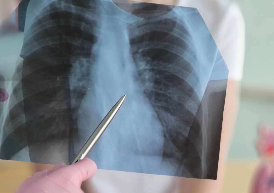
A lung nodule is a well-defined abnormality within the lung, usually detected on a chest X-ray or computed tomography (CT) scan. It can arise from a variety of causes, such as past infections, scar tissue, inflammatory diseases, benign growths or lung cancer.

Finding a nodule in the lung does not, on its own, mean a diagnosis of cancer.

When a nodule is evaluated, the assessment looks not only at its size but also at its shape, margins, density, location within the lung and whether it has changed compared with previous imaging. The person's age, smoking history, any prior history of cancer and other individual risk factors are also taken into account.

Based on this evaluation, imaging follow-up may be sufficient for some nodules, while others may require PET-CT, biopsy or surgical evaluation.

What Is a Lung Nodule?

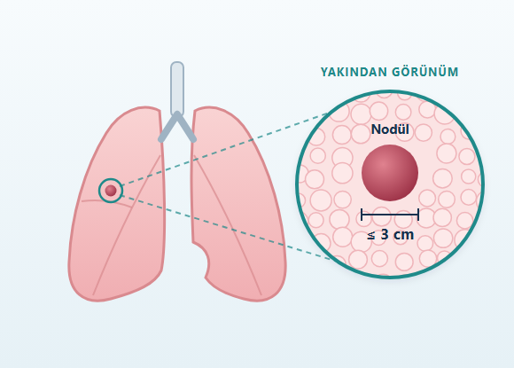
A lung nodule is generally defined as a well-defined abnormality up to 3 centimeters in diameter that can be distinguished from the surrounding normal lung tissue. Larger lesions are usually referred to as a lung mass.

Lung nodules may:
  • Be single
  • Be multiple
  • Be solid
  • Have a ground-glass appearance
  • Contain both ground-glass and solid areas


Some nodules are found incidentally on a CT scan performed for another reason. Others are discovered while investigating respiratory symptoms or another lung disease.

Does a Lung Nodule Cause Symptoms?

Small lung nodules usually do not cause any symptoms. For this reason, a large proportion of nodules are found incidentally during imaging performed for another reason.

A person may also have symptoms such as:
  • Cough
  • Shortness of breath
  • Chest pain
However, the presence of these symptoms does not necessarily mean that the nodule is causing them. Other diseases affecting the respiratory system can cause similar symptoms.

What Causes a Lung Nodule?

There is no single cause of lung nodules.

Possible causes include:
  • Previous lung infections
  • Scar tissue in the lung
  • Tuberculosis and other granulomatous diseases
  • Certain fungal infections
  • Benign lung tumors
  • Inflammatory diseases
  • Certain rheumatologic diseases
  • Primary lung cancer
  • Spread of cancer from another organ to the lung
The cause of a nodule cannot always be determined with certainty from the CT image alone. For this reason, imaging features, changes over time and the person's risk profile are evaluated together.

What Are the Types of Lung Nodules?

Lung nodules can be divided into different groups based on their density and structural features on computed tomography. This distinction is important because it can affect how the nodule is followed and evaluated.

What Is a Solid Lung Nodule?

A solid nodule is a lesion that appears denser than the surrounding lung tissue on CT. Solid nodules can develop from both benign and malignant causes.

The evaluation takes into account features such as:
  • Size
  • Shape
  • Margins
  • Location within the lung
  • Growth compared with previous imaging

What Is a Ground-Glass Nodule?

A ground-glass nodule appears on CT as a hazy area of increased density that does not completely obscure the underlying lung structures. Temporary ground-glass areas can form due to infection or inflammation. However, some ground-glass nodules may persist.

In persistent ground-glass nodules, the following are important:
  • The size of the nodule
  • Its appearance
  • Its change over time
  • Whether a new solid area has developed
Depending on the nodule's characteristics, longer-term imaging follow-up or further evaluation may be needed.

What Is a Part-Solid Nodule?

Part-solid nodules contain both a ground-glass component and a denser solid component. In these nodules, the size of the solid portion and its change over time are particularly important. Growth of the solid component or an increase in its density may affect the need for further investigation.

Is a Lung Nodule Cancer?

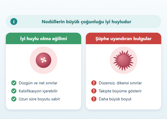
Not every lung nodule is cancer. A large proportion of lung nodules arise from non-cancerous causes. However, some nodules may be the first imaging finding of early-stage lung cancer. For this reason, the key question is not simply "Is there a nodule?" The likelihood that the nodule is cancerous needs to be assessed.
How Is the Cancer Risk of a Lung Nodule Assessed?

No single feature definitively shows whether a nodule is benign or malignant.

The evaluation may consider the following together:
  • The size of the nodule
  • Its shape
  • Its margins
  • Whether it is solid, ground-glass or part-solid
  • Its location within the lung
  • Growth compared with previous imaging
  • The person's age
  • Smoking history
  • Occupational and environmental exposures
  • Prior history of cancer
  • Family history of lung cancer
A small nodule should not automatically be considered harmless simply because it is small. Likewise, a larger nodule does not, on its own, mean a diagnosis of cancer.

Why Are Previous CT Scans Important in Evaluating a Lung Nodule?

Comparing the current CT with previous imaging can provide important information when evaluating a nodule. A nodule that has remained unchanged for a long time is not evaluated in the same way as a nodule that is new or has grown over time.

During the comparison, the following may be examined:
  • Change in diameter
  • Change in volume
  • Change in density
  • Development of a solid component
  • Changes in margins or shape
For this reason, previous chest X-rays or CT images can be important for the evaluation.

How Is a Lung Nodule Followed Up?

The duration of follow-up and the imaging interval are not the same for every lung nodule.

When planning follow-up, the following are evaluated together:
  • The size of the nodule
  • The type of nodule
  • Imaging features
  • Changes compared with previous imaging
  • The person's risk factors for cancer
For some nodules, CT follow-up at regular intervals may be sufficient. The goal of follow-up is not only to measure the diameter of the nodule. Changes in features such as density, shape and the solid component are also evaluated.

Why Is Computed Tomography Important for a Lung Nodule?

Computed tomography is one of the main imaging methods for evaluating a lung nodule in detail.

CT can be used to evaluate:
  • The size of the nodule
  • Its density
  • Its shape
  • Its margins
  • Its location
  • Its relationship with surrounding structures
In addition, the behavior of the nodule over time can be examined by comparing earlier and later CT scans.

When Is PET-CT Used for a Lung Nodule?

PET-CT is an imaging method that evaluates the metabolic activity of tissues. It may be used in the further evaluation of some lung nodules or in investigating a suspicion of lung cancer. However, PET-CT has important limitations. Increased uptake on PET-CT does not, on its own, establish a diagnosis of cancer. Infections and inflammatory diseases can also show increased metabolic activity.

Conversely:
  • Very small nodules
  • Some tumors with low metabolic activity
may not show significant uptake on PET-CT. For this reason, PET-CT results are evaluated together with other imaging findings and, when necessary, pathological examination.

When Might a Biopsy Be Needed for a Lung Nodule?

Not every lung nodule is biopsied.

When assessing the need for a biopsy, the following are evaluated together:
  • The size of the nodule
  • Imaging features
  • Whether it shows growth
  • Its location
  • The estimated likelihood of cancer
  • The technical feasibility of a biopsy
  • The potential risks of the procedure
Obtaining a reliable tissue sample from very small nodules can be technically difficult. In some cases, the risk of the biopsy may be higher than the estimated risk of the nodule. For this reason, imaging follow-up may be preferred in suitable patients.

Can a Lung Nodule Be Biopsied with Bronchoscopy?

Bronchoscopy is a method that allows the trachea and bronchi to be examined with the help of a camera. It can be used particularly to obtain tissue samples from certain nodules or masses located near the central airways. The location of the nodule and its relationship with the airways can affect whether a bronchoscopic biopsy is appropriate. It may not be possible to reach every peripheral lung nodule with standard bronchoscopy.

What Is EBUS and Why May It Be Used for a Lung Nodule?

Endobronchial ultrasound, or EBUS, is a method that combines bronchoscopy with ultrasound imaging. It may be used particularly to evaluate certain lymph nodes within the chest and to obtain tissue samples from these areas. When there is a suspicious lung nodule or mass together with abnormal lymph nodes, EBUS may be part of the diagnostic and possible staging process.

What Is a Transthoracic Needle Biopsy?

For some nodules located near the outer regions of the lung, a tissue sample can be obtained under CT guidance with a needle passed through the chest wall. This method is called a transthoracic needle biopsy. It is not suitable for every nodule.

Factors that may affect suitability include:
  • The size of the nodule
  • The location of the nodule
  • Its distance from the chest wall
  • Nearby blood vessels
  • The person's existing lung structure
Possible complications include pneumothorax and bleeding.

What Is Multidisciplinary Evaluation in Lung Cancer?

The diagnosis and treatment of lung cancer may require joint evaluation by different medical specialties.

Specialties such as the following may be involved in the process:
  • Thoracic Surgery
  • Pulmonology
  • Medical oncology
  • Radiation oncology
  • Radiology
  • Nuclear medicine
  • Pathology
This approach can be particularly important for confirming the diagnosis, ensuring accurate staging and determining the order in which different treatment options are applied.

What Is a Surgical Biopsy?

A surgical biopsy may be considered when a diagnosis cannot be made with other biopsy methods, or when the imaging features suggest that surgical removal of the nodule may be appropriate. The nodule can be removed together with a limited amount of surrounding lung tissue. In some cases, this procedure can be a step toward both diagnosis and treatment planning.

The extent of the operation may vary depending on:
  • The location of the nodule
  • Its size
  • Findings during surgery
  • Pathological evaluation

When Is a Lung Nodule Operated On?

Finding a lung nodule does not, on its own, require surgery.

Surgical evaluation may be considered particularly in situations such as:
  • Growth of the nodule over time
  • The presence of suspicious imaging features
  • The appearance or growth of a solid component
  • A significant likelihood of cancer
  • Inability to make a diagnosis with other biopsy methods
  • Surgical removal of the nodule being considered appropriate for diagnosis and treatment
The decision to operate is not based on the size of the nodule alone.

How Much Lung Tissue Is Removed in Lung Nodule Surgery?

The amount of lung tissue to be removed may vary depending on the characteristics of the nodule and the purpose of the surgery.

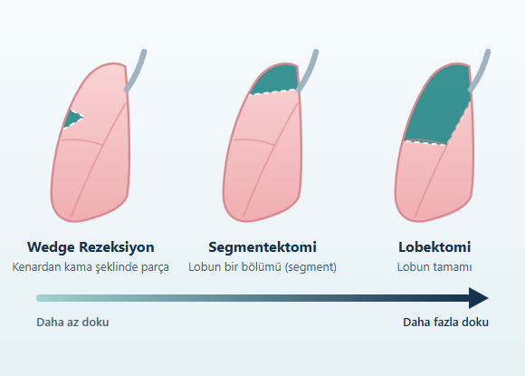
Wedge Resection

The nodule is removed together with a limited amount of surrounding lung tissue. A wedge resection does not follow the anatomical segment boundaries of the lung and may be used for diagnostic purposes in some nodules.

Segmentectomy

This is the removal of an anatomical segment of a lung lobe together with the bronchus and blood vessels belonging to that segment. When the nodule is found to be lung cancer, or in selected cases where the likelihood of cancer is high, an anatomical segmentectomy may be considered depending on the characteristics of the tumor. More extensive lung cancer operations are planned separately according to the type, stage and anatomical features of the cancer.
Can a Lung Nodule Be Removed with VATS?

In suitable patients, surgical evaluation and removal of lung nodules can be performed with video-assisted thoracoscopic surgery (VATS). VATS is a minimally invasive surgical method performed through small incisions in the chest wall using a camera and special surgical instruments.

Procedures that can be performed with VATS include:
  • Surgical lung biopsy
  • Nodule removal
  • Wedge resection
  • Segmentectomy
However, suitability for a minimally invasive approach is assessed based on the size and location of the nodule, its relationship with surrounding structures, adhesions within the chest and the extent of the planned operation.

What Is the Difference Between a Lung Nodule and Lung Cancer?

A lung nodule is an imaging finding. Lung cancer, on the other hand, is a disease defined by its cellular and pathological characteristics. The appearance of a nodule on CT can provide important information about the likelihood of cancer, but not every nodule can be definitively classified as benign or malignant by imaging. Some nodules may remain unchanged for years.

Others may:
  • Grow
  • Change in density
  • Change in shape
  • Develop a new solid component
Proper evaluation of a suspicious lung nodule may allow some lung cancers to be detected while still at an early and limited stage. On the other hand, avoiding unnecessary biopsies and surgical procedures for low-risk nodules is also an important part of proper nodule management.

Frequently Asked Questions About Lung Nodules

Does having a nodule in my lung mean I have cancer?
No. A large proportion of lung nodules arise from benign causes. The nodule's size, shape, density, change over time and the person's individual cancer risk factors are evaluated together.
What size lung nodule is dangerous?
There is no single size threshold that can classify a nodule as dangerous or harmless based on size alone. Larger nodules may generally require more detailed evaluation, but size should be assessed together with the nodule's structure, density, growth and the person's risk factors.
Can a lung nodule go away on its own?
Some nodules that develop due to infection or inflammation may shrink or disappear completely over time. For persistent nodules, imaging follow-up or further evaluation may be needed depending on their characteristics.
Is a ground-glass nodule cancer?
Not every ground-glass nodule is cancer. Infections and inflammatory diseases can also cause a ground-glass appearance. However, some ground-glass nodules that persist or change over time may be associated with early-stage lung tumors.
If a lung nodule grows, is it cancer?
Growth of a nodule is an important finding in the evaluation, but it does not, on its own, establish a definitive diagnosis of cancer. The growth rate, structure, density and shape of the nodule and other risk factors are evaluated together.
Does uptake in a nodule on PET-CT definitely mean cancer?
No. Infections and inflammatory diseases can also show increased metabolic activity on PET-CT. Some cancers, on the other hand, may show low or no significant uptake.
Is every lung nodule biopsied?
No. Some nodules are followed with imaging, while others may require biopsy or surgical evaluation. The decision is based on the characteristics of the nodule and the person's risk profile.
How often is a lung nodule followed up?
The follow-up interval is not the same for every nodule. The size of the nodule, whether it is solid or subsolid, its imaging features, changes compared with previous scans and the person's risk factors all affect the follow-up plan.
Can a lung nodule be removed with surgery?
In appropriate cases, the nodule can be surgically removed. Surgery may be considered to obtain a diagnosis or to appropriately remove a lesion suspected of being cancer.
Can a lung nodule be removed with minimally invasive (closed) surgery?
In suitable patients, nodule removal, wedge resection or segmentectomy can be performed using minimally invasive methods such as VATS. The method to be used is determined according to the location and characteristics of the nodule and the extent of the planned surgery.

Conclusion

A lung nodule is not, on its own, a diagnosis of cancer. Nodules can arise from a variety of causes, ranging from past infections and benign growths to lung cancer. For this reason, the evaluation is not based on the size of the nodule alone. The nodule's:
  • Solid, ground-glass or part-solid structure
  • Size
  • Shape and margins
  • Location within the lung
  • Change over time
are evaluated together with the person's individual risk factors. For some nodules, CT follow-up at regular intervals may be sufficient. Others may require PET-CT or biopsy. In selected cases, surgical biopsy or removal of the nodule may be considered. The main goal is to evaluate suspicious nodules appropriately while avoiding unnecessary interventions for nodules that carry a low risk of cancer.

Disclaimer: The information on this page is provided for general informational purposes only and does not replace a medical examination, diagnosis or an individualized follow-up and treatment plan. Decisions about follow-up, further imaging, biopsy or surgical evaluation for lung nodules are made individually, based on the nodule's size, structure, imaging features and change over time, as well as the person's risk factors and overall health.