Pleura
Information on Pleural Effusion, Empyema, Pleural Infection, Malignant Pleural Effusion, Mesothelioma, Chylothorax, and the Treatment of Pleural Diseases.
Pleural Diseases
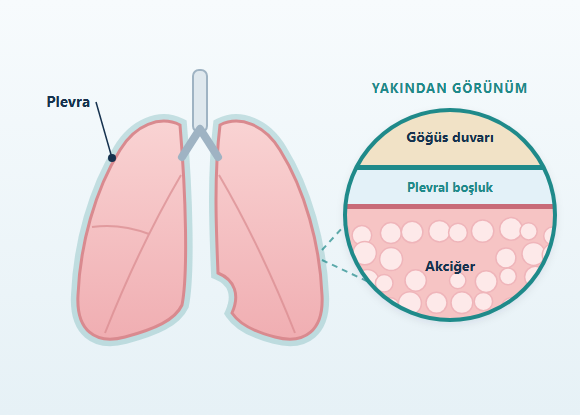
The pleura is a thin, two-layered membrane that covers the outer surface of the lungs and lines the inside of the chest cavity. The layer covering the lung surface is called the visceral pleura, and the layer lining the inner surface of the chest wall is called the parietal pleura.
Between these two layers is a narrow space called the pleural space. Under normal conditions, this space contains a small amount of fluid that allows the lungs to move freely within the rib cage during breathing.
An abnormal buildup of fluid, air, blood, lymph, or infected material in the pleural space can lead to various pleural diseases. Inflammation, thickening, or tumor involvement of the pleural tissue can also affect lung expansion and respiratory function.
The main pleural diseases include:
- Pleural effusion
- Parapneumonic effusion
- Pleural infection and empyema
- Malignant pleural effusion
- Pleural tumors
- Pleural mesothelioma
- Chylothorax
- Hemothorax
- Pleural thickening
- Non-expandable lung
What Are the Symptoms of Pleural Diseases?
Symptoms may vary depending on the type of pleural disease, the amount of fluid or other material that has accumulated in the pleural space, and the underlying cause.
Possible symptoms include:
- Shortness of breath
- Chest pain
- Stabbing pain that worsens with deep breathing
- Cough
- Fever
- Chills and shivering
- Fatigue
- Reduced exercise capacity
- Loss of appetite
- Unintentional weight loss
- A feeling of heaviness or pressure in the chest
What Is Pleural Effusion?
Pleural effusion is an abnormal buildup of fluid in the pleural space between the lung and the chest wall.
It is sometimes described in everyday language as "water on the lung." However, the fluid is not inside the lung tissue itself but in the pleural space surrounding the lung.
Pleural effusion is not a disease in itself.
It is a finding that can result from various conditions affecting the heart, lungs, kidneys, liver, or pleura.
The fluid may be:
- One-sided
- Two-sided
- Free-flowing
- Loculated (divided into pockets)
What Causes Pleural Effusion?
Causes of pleural fluid buildup may include:
- Heart failure
- Pneumonia and other lung infections
- Lung cancer
- Tumors that have spread to the pleura from other organs
- Pleural mesothelioma
- Pulmonary embolism
- Kidney diseases
- Liver cirrhosis
- Tuberculosis
- Rheumatologic diseases
- Pancreatic diseases
- Chest trauma
- Heart or thoracic surgery
- Certain medications
- Lymphatic circulation disorders
What Are Transudate and Exudate?
Pleural fluids are generally classified into two groups, transudative and exudative, based on the mechanism by which they form.
This distinction can be made by evaluating biochemical values such as protein and LDH in pleural fluid and blood samples together.
Transudative Pleural Effusion
A transudative effusion usually results from changes in the body's fluid pressures or protein balance.
Common causes include:
- Heart failure
- Liver cirrhosis
- Certain kidney diseases
Exudative Pleural Effusion
An exudative effusion may develop as a result of processes in the pleura or surrounding tissues, such as:
- Infection
- Inflammation
- Increased vascular permeability
- Tumor involvement
How Is Pleural Effusion Evaluated?
The evaluation takes the following into account together:
- The person's complaints
- Medical history
- Physical examination
- Imaging results
- Pleural fluid analysis, when necessary
- Pleural biopsy, when needed

Chest X-Ray
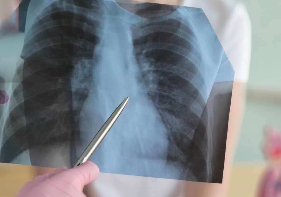
A chest X-ray is one of the basic imaging methods used to detect fluid in the pleural space.
It can provide general information about the amount of fluid and its effect on the lung.
Small amounts of pleural fluid may not always be visible on a standard chest X-ray.
Chest Ultrasound
Ultrasound helps evaluate:
- The amount of pleural fluid
- Its location
- Whether it is free-flowing or loculated
Computed Tomography
Computed tomography (CT) shows the lungs, pleura, chest wall, and mediastinum in detail.
CT can be used to evaluate:
- The distribution of pleural fluid
- Pleural thickening
- Pleural nodules or masses
- Lung tumors
- Lung infection
- Lung abscess
- Lymph nodes
- Chest wall involvement
- The ability of the lung to expand
What Is Thoracentesis?
Thoracentesis is a procedure in which fluid that has accumulated in the pleural space is removed using a needle or a thin catheter.
It can be performed for two main purposes:
- To obtain a sample to investigate the cause of the fluid
- To drain excess fluid and relieve shortness of breath
- Protein and other biochemical values
- Cell count
- Bacteria and other microorganisms
- Tuberculosis-related tests
- Cancer cells
- Fat content
- Blood content
- Pain
- Bleeding
- Infection
- Pneumothorax
When Is a Pleural Biopsy Performed?
Laboratory analysis of pleural fluid does not always lead to a definitive diagnosis.
A tissue sample may be needed if the pleura shows:
- Thickening
- A nodule
- A mass
- Unexplained one-sided fluid
- Suspected tumor
- Image-guided needle biopsy
- Medical thoracoscopy
- Video-assisted thoracoscopic surgery (VATS)
Thoracoscopy and VATS
Thoracoscopy is the general term for methods that allow the pleural space to be examined directly with a camera.
VATS is a minimally invasive thoracic surgery technique performed with a camera and surgical instruments through small incisions in the chest wall.
Procedures that can be performed with VATS include:
- Pleural biopsy
- Drainage of pleural fluid
- Removal of infected material
- Release of pleural adhesions
- Pleurodesis
- Evaluation of pleural tumors
- Decortication
What Is Parapneumonic Effusion?
A buildup of pleural fluid that accompanies pneumonia is called a parapneumonic effusion.
Some parapneumonic effusions may resolve with appropriate antibiotic treatment.
However, drainage may be needed if the fluid becomes infected, its chemical characteristics change, it becomes divided into pockets, or it prevents the lung from expanding.
Clinically, different stages may be seen:
- Simple parapneumonic effusion
- Complicated parapneumonic effusion
- Empyema
What Is Empyema?
Empyema is the presence of infected fluid or pus in the pleural space.
Although it is most often associated with pneumonia, it can also develop after:
- Chest surgery
- Chest trauma
- Injury to the esophagus
- Infections in other parts of the body
- Fever
- Chills and shivering
- Chest pain
- Cough
- Shortness of breath
- Fatigue
- Loss of appetite
- Night sweats
- Unintentional weight loss
How Is Empyema Treated?
The main goals of treating pleural infection and empyema are:
- Bringing the infection under control
- Adequately draining the infected pleural fluid
- Allowing the lung to re-expand whenever possible.
- Antibiotics
- Chest tube drainage
- Medications delivered into the pleural space in suitable patients
- VATS
- Decortication
Antibiotic Treatment
The choice of antibiotic is based on factors such as:
- Whether the infection was acquired in the community or in the hospital
- Antibiotics used previously
- Coexisting conditions
- Culture results
Chest Tube Drainage
In pleural infection, a chest tube may be used to drain the infected fluid.
Drainage may be more difficult when the fluid is divided into multiple pockets.
The position of the chest tube and the adequacy of drainage can be assessed with ultrasound or computed tomography.
Medications Delivered into the Pleural Space
If loculated or residual infected fluid remains in the pleural space despite initial chest tube drainage, treatment with medications delivered into the pleural space may be considered in selected patients.
For this purpose, a combination of tissue plasminogen activator (tPA) and DNase may be used.
The aim is to break down fibrin structures, help thick infected material flow more easily, and improve drainage.
This treatment is not suitable for every patient.
Factors such as bleeding risk and use of blood-thinning medications are evaluated before treatment.
VATS Debridement
Surgical treatment may be considered in some patients whose infection cannot be adequately controlled despite chest tube drainage and appropriate medical therapy. With VATS:- Infected fluid can be drained
- The pleural space can be cleaned out
- Fibrin and infected tissue can be removed
- Adhesions preventing lung expansion can be released
Decortication
In advanced pleural infection, a thick, rigid fibrous layer may develop on the outer surface of the lung. If this layer prevents the lung from re-expanding, decortication may be considered. Decortication is the surgical removal of this thick tissue surrounding the lung. Not every patient with empyema requires decortication. The decision is made by taking into account:- The stage of the infection
- The ability of the lung to expand
- Symptoms
- The person's physical condition and fitness for surgery
What Is Malignant Pleural Effusion?
Malignant pleural effusion is a buildup of fluid that develops when the pleural fluid or pleural tissue is involved by cancer.
Lung cancer, breast cancer, lymphoma, and tumors of various other organs can spread to the pleura.
Pleural mesothelioma can also cause pleural fluid buildup.
Symptoms may include:
- Shortness of breath
- Heaviness in the chest
- Cough
- Chest pain
- Reduced exercise capacity
How Is Malignant Pleural Effusion Treated?
Treatment is not based solely on how much space the fluid occupies on imaging.
Planning takes the following into account together:
- The type of cancer
- The systemic treatment being given
- How quickly the fluid re-accumulates
- The degree of shortness of breath
- Whether the lung can expand after the fluid is drained
- The person's overall health
- The expected course of treatment
- Previous pleural procedures
- Thoracentesis
- Chest tube
- Pleurodesis
- Indwelling pleural catheter
- Thoracoscopic procedures
- Systemic treatments for the underlying cancer
What Is Pleurodesis?
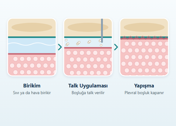
Pleurodesis is a procedure that aims to eliminate the pleural space by causing the visceral and parietal pleural layers to adhere to each other in a controlled manner.
This is intended to reduce the likelihood of fluid re-accumulating in the same space.
Pleurodesis can be performed:
- Through a chest tube
- During thoracoscopy
What Is an Indwelling Pleural Catheter (IPC)?
An indwelling pleural catheter is a tunneled drainage system that allows recurrent pleural fluid to be drained at regular intervals without repeated procedures in the hospital. It may be considered particularly:- In recurrent symptomatic malignant pleural effusion
- In some cases where the lung cannot expand adequately
- Infection
- Blockage
- Fluid leakage around the catheter
- Pain
- Displacement
- Pleural infection
What Is a Non-Expandable Lung?
When the lung cannot expand sufficiently toward the chest wall after the pleural fluid is drained, this is referred to as a non-expandable lung. This condition can develop due to causes such as:- Tumor involvement of the pleura
- Long-standing pleural infection
- Pleural thickening
- Loss of elasticity in the lung tissue
- Blockage of one of the main airways
Pleural Tumors
Pleural tumors can be benign or malignant. Some tumors arise directly from the pleural tissue, while cancers originating in the lung, breast, and other organs can also spread to the pleura. Imaging may show:- Pleural thickening
- Pleural nodules
- A pleural mass
- Recurrent pleural fluid
- Chest wall involvement
What Is Pleural Mesothelioma?
Pleural mesothelioma is a malignant tumor that develops from the mesothelial cells of the pleura. Asbestos exposure is one of the most important risk factors associated with the disease. However, not everyone exposed to asbestos develops mesothelioma, and in some patients no clear history of exposure can be identified. Many years may pass between asbestos exposure and the onset of the disease. Symptoms may include:- One-sided chest pain
- Shortness of breath
- Recurrent pleural fluid
- Cough
- Unintentional weight loss
- Fatigue
- A feeling of pressure in the chest
- Pleural thickening
- Nodular pleural structures
- Pleural fluid
- Involvement of the chest wall or other neighboring structures
How Is Pleural Mesothelioma Treated?
Treatment of pleural mesothelioma is planned in a multidisciplinary manner, taking into account:
- The stage and extent of the disease
- The histologic subtype
- Whether it can be surgically removed
- The person's respiratory capacity
- Overall health
- Coexisting conditions
- Systemic drug therapies
- Immunotherapy
- Chemotherapy
- Radiation therapy
- Procedures to control pleural fluid
- Surgical treatment in selected patients
What Is Chylothorax?
Chylothorax is the buildup in the pleural space of a fluid called chyle, which is carried within the lymphatic system.
Chylothorax can develop as a result of:
- Thoracic surgery
- Trauma
- Lymphoma and other tumors
- Diseases of the lymphatic ducts
- Certain congenital disorders
- Dietary changes
- Intravenous nutrition in selected cases
- Medical treatments aimed at reducing lymph flow
- Pleural drainage
- Interventional radiology procedures
- Surgical procedures targeting the thoracic duct
What Is Hemothorax?
Hemothorax is a buildup of blood in the pleural space.
Causes may include:
- Chest trauma
- Rib fractures
- Injuries to the lung or chest wall
- Medical procedures
- Thoracic surgery
- Blood vessel injuries
- Certain tumors
What Is Pleural Thickening?
Pleural thickening means that the pleural tissue appears thicker than normal.
Causes may include:
- Previous infection
- Pleural bleeding
- Asbestos exposure
- Surgery
- Trauma
- Inflammatory diseases
- Tumors
- Whether the thickening is smooth or nodular
- Whether it is on one or both sides
- Which parts of the pleura are affected
- Its relationship to surrounding organs
- How it has changed compared with previous imaging
How Is Treatment Determined in Pleural Diseases?
Treatment is not determined solely by the amount of fluid in the pleural space. Planning takes the following into account together:- The cause of the fluid
- The presence of infection
- The presence of a tumor
- How quickly the fluid recurs
- The ability of the lung to expand
- The degree of shortness of breath
- Overall health
- Respiratory and cardiac capacity
- Previous procedures
- Surgical risk
A Multidisciplinary Approach to Pleural Diseases
The cause of pleural disease may be:- Infection
- Cancer
- Heart disease
- Rheumatologic disease
- Systemic diseases
- Trauma
- Surgical complications
- Thoracic surgery
- Pulmonology
- Radiology
- Pathology
- Infectious diseases
- Medical oncology
- Radiation oncology
- Cardiology
- Rheumatology
Follow-Up After Treatment
The follow-up plan is determined by the cause of the pleural disease and the treatment given. During follow-up, the following may be evaluated:- The course of shortness of breath
- Whether pleural fluid has re-accumulated
- The expansion of the lung
- Signs of infection
- The function of the chest tube or catheter
- The surgical wound
- Pathology results
- The course of the underlying disease
Frequently Asked Questions About Pleural Diseases
What does "water on the lung" mean?
The condition commonly referred to as "water on the lung" is usually a pleural effusion. The fluid does not collect inside the lung tissue but in the pleural space between the lung and the chest wall.
Does pleural effusion mean cancer?
No. Heart failure, infection, kidney or liver disease, pulmonary embolism, and rheumatologic diseases can also cause pleural effusion. Cancer is only one of the possible causes.
Does pleural fluid always need to be drained?
No. The decision is based not only on the amount of fluid but also on its cause and the person's symptoms and clinical condition. Some small effusions can be monitored, while fluid that is infected or causes significant shortness of breath may require drainage.
Does thoracentesis completely cure pleural effusion?
Thoracentesis makes it possible to investigate the cause of the fluid and, in some patients, to relieve shortness of breath. However, treatment of the underlying disease or additional pleural procedures may be needed to prevent the fluid from re-accumulating.
Does the absence of cancer cells in pleural fluid completely rule out cancer?
No. Pleural fluid cytology may not reveal cancer cells in every pleural tumor. If imaging findings or clinical suspicion persist, a pleural biopsy may be needed.
Does empyema always require surgery?
No. Treatment usually begins with antibiotics and appropriate drainage. In selected patients in whom drainage is inadequate, medications delivered into the pleural space or surgical treatment may be considered.
When is pleurodesis used?
Pleurodesis can be used particularly to control recurrent symptomatic pleural fluid. For the procedure to be effective, it is important that the lung can expand sufficiently after the fluid is drained.
Does an indwelling pleural catheter stay in place for life?
Not always. How long it is used can vary depending on whether the fluid continues to re-accumulate, whether catheter drainage continues, and, in some patients, whether the pleural layers adhere on their own over time.
Is pleural thickening cancer?
No. Pleural thickening can develop for various reasons, such as infection, trauma, surgery, bleeding, and asbestos exposure. Further evaluation may be needed for thickening that is nodular, progressive, or unexplained.
Does everyone exposed to asbestos develop mesothelioma?
No. Asbestos is an important risk factor for pleural mesothelioma, but not everyone exposed to asbestos develops mesothelioma. In some people, the disease can occur without a clear history of asbestos exposure.
Disclaimer: The information on this website is intended for general informational purposes only and is not a substitute for a personal medical evaluation. In pleural diseases, decisions about diagnosis, follow-up, and treatment are made individually by the physician based on the person's clinical findings, imaging results, pleural fluid and, when necessary, tissue analyses, and overall health.